9NYS | pdb_00009nys

Human DNA Ligase 1 E346A/E592A/K845N triple mutant with 3'-A:T nick


Experimental Data Snapshot

  • Method: X-RAY DIFFRACTION
  • Resolution: 2.64 Å
  • R-Value Free: 
    0.223 (Depositor), 0.222 (DCC) 
  • R-Value Work: 
    0.185 (Depositor), 0.187 (DCC) 
  • R-Value Observed: 
    0.188 (Depositor) 

Starting Model: experimental
View more details

wwPDB Validation   3D Report Full Report


Ligand Structure Quality Assessment 


This is version 1.0 of the entry. See complete history


Literature

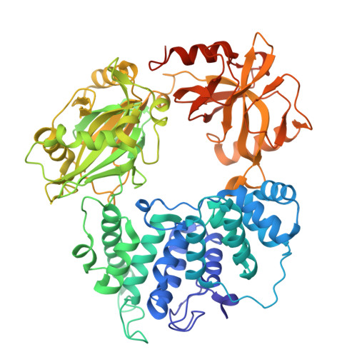
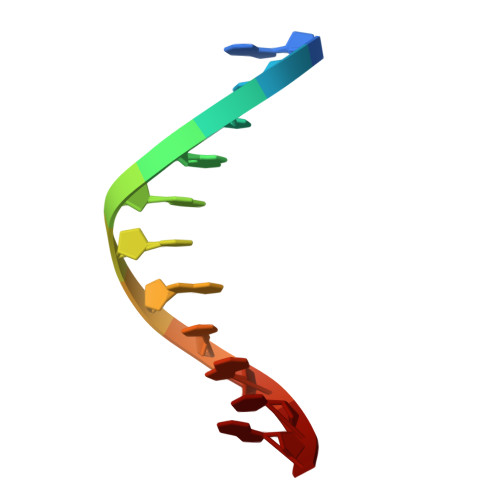
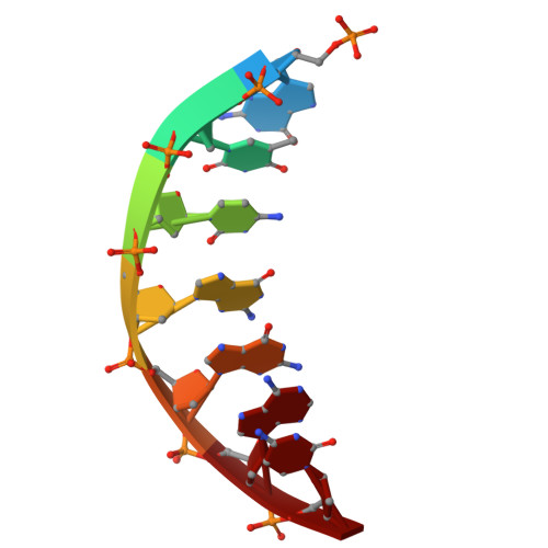
Impaired nick recognition and ligation efficiency by LIG1 K845N variant linked to Huntington's disease.

Ratcliffe, J.Lerner, C.E.Balu, K.Chatterjee, S.Lee, K.M.Caglayan, M.

(2025) NAR Mol Med 2: ugaf038-ugaf038

  • DOI: https://doi.org/10.1093/narmme/ugaf038
  • Primary Citation of Related Structures:  
    9NYS

  • PubMed Abstract: 

    DNA ligase 1 (LIG1) joins broken strand breaks and seals Okazaki fragments during DNA repair and replication. Huntington's disease (HD)-associated mutation in the  LIG1 gene, K845N, is associated with delayed symptom onset and predicted to suppress CAG repeat expansion. Yet, how this mutation impacts faithful nick sealing and efficient DNA binding by LIG1 remains unknown. Here, using biochemical analyses, X-ray crystallography, and total internal reflection fluorescence (TIRF) microscopy, we characterized the LIG1 HD-associated K845N variant at biochemical, structural, and single-molecule levels. Our results showed significantly reduced ligation efficiency for nick substrates containing noncanonical mismatches and diminished mutagenic end-joining of damaged DNA, while LIG1 K845N variant exhibits a lack of discrimination against nicks containing 3'-ribonucleotides when compared with the wild-type enzyme. Furthermore, our structures provided an atomic insight into differences in the distances between functional groups of K/N845 and DNA ends, demonstrating similar conformation at the ligase active site. Finally, our single-molecule measurements revealed that the K845N variant binds less frequently to nick, suggesting diminished affinity. Overall, our findings contribute to understanding the mechanism by which LIG1 searches for nick sites on DNA and ensures fidelity to maintain genome stability at the final ligation step in normal versus HD-associated states.


  • Organizational Affiliation
    • Department of Biochemistry and Molecular Biology, University of Florida, Gainesville, FL 32610, United States.

Macromolecules

Find similar proteins by:  (by identity cutoff)  |  3D Structure
Entity ID: 1
MoleculeChains Sequence LengthOrganismDetailsImage
DNA ligase 1668Homo sapiensMutation(s): 3 
Gene Names: LIG1
EC: 6.5.1.1
UniProt & NIH Common Fund Data Resources
Find proteins for P18858 (Homo sapiens)
Explore P18858 
Go to UniProtKB:  P18858
PHAROS:  P18858
GTEx:  ENSG00000105486 
Entity Groups  
Sequence Clusters30% Identity50% Identity70% Identity90% Identity95% Identity100% Identity
UniProt GroupP18858
Sequence Annotations
Expand
  • Reference Sequence

Find similar nucleic acids by:  Sequence   |   3D Structure  

Entity ID: 2
MoleculeChains LengthOrganismImage
DNA (5'-D(*GP*CP*TP*GP*AP*TP*GP*CP*GP*TP*A)-3')11synthetic construct
Sequence Annotations
Expand
  • Reference Sequence

Find similar nucleic acids by:  Sequence   |   3D Structure  

Entity ID: 3
MoleculeChains LengthOrganismImage
DNA (5'-D(P*GP*TP*CP*GP*GP*AP*C)-3')7synthetic construct
Sequence Annotations
Expand
  • Reference Sequence

Find similar nucleic acids by:  Sequence   |   3D Structure  

Entity ID: 4
MoleculeChains LengthOrganismImage
DNA (5'-D(*GP*TP*CP*CP*GP*AP*CP*TP*AP*CP*GP*CP*AP*TP*CP*AP*GP*C)-3')18synthetic construct
Sequence Annotations
Expand
  • Reference Sequence
Experimental Data & Validation

Experimental Data

  • Method: X-RAY DIFFRACTION
  • Resolution: 2.64 Å
  • R-Value Free:  0.223 (Depositor), 0.222 (DCC) 
  • R-Value Work:  0.185 (Depositor), 0.187 (DCC) 
  • R-Value Observed: 0.188 (Depositor) 
Space Group: P 21 21 21
Unit Cell:
Length ( Å )Angle ( ˚ )
a = 74.668α = 90
b = 100.123β = 90
c = 115.089γ = 90
Software Package:
Software NamePurpose
PHENIXrefinement
XDSdata reduction
XDSdata scaling
PHASERphasing

Structure Validation

View Full Validation Report


Ligand Structure Quality Assessment 


Entry History & Funding Information

Deposition Data


Funding OrganizationLocationGrant Number
National Institutes of Health/National Institute of General Medical Sciences (NIH/NIGMS)United States1R35GM14711101

Revision History  (Full details and data files)

  • Version 1.0: 2025-12-31
    Type: Initial release