Block of Kv1 potassium channels and NMR structure of recombinant conotoxin kappa-SrXIA.
Escobar, L.I., Quezada Suaste, C.D., Salvador, C., Aparicio, D.L., Melchor-Meneses, C.M., Bravo-Martinez, J., de la Rosa, V., Lopez-Gonzalez, Z., Del Rio-Portilla, F.(2025) Toxicon 262: 108384-108384
- PubMed: 40324600 Search on PubMed
- DOI: https://doi.org/10.1016/j.toxicon.2025.108384
- Primary Citation Related Structures:
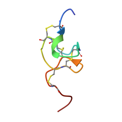
9NVH - PubMed Abstract:
The biologically active components of Conus venoms are mainly small peptides with disulfide-bonded structures. Some conotoxins contain post-translational modifications as an evolutionary strategy to enhance their potency and selectivity towards ion channels and receptors. Few conotoxins are known to target mammalian Kv1 channels. κ-SrXIA from the venom of Conus spurius inhibits the voltage-gated potassium Kv1.2 and Kv1.6 channels through a basic ring of Arg. The 32 amino acid κ-SrXIA has eight Cys residues arranged in the pattern that defines the I-superfamily with four disulfide bridges, two gamma-carboxy-glutamates (Gla), and a Pro-amidated C-terminus (X). In this study, we obtained and determined the biological activity and NMR structure of recombinant κ-SrXIA without Gla and X. The conotoxin cDNA was expressed in E. coli CD41 and purified by GST-affinity chromatography and RP-HPLC. Pharmacological assays were performed by two-electrode voltage-clamp recordings in Xenopus laevis oocytes expressing recombinant Kv1.1, Kv1.2, Kv1.3, Kv1.4 and Kv1.6 channels. Except for Kv1.3, κ-Sr-XIA irreversibly blocked Kv1 channels displaying a lower affinity and a slower inhibition kinetics than native conotoxin. Even when κ-SrXIA displayed a high structural similarity to GXIA from the I 3 -superfamily, the resultant disulfide connectivity forming an ICK+1 motif showed two antiparallel β-strands as ι-RXIA from the I 1 -superfamily. This study represents the first 3D NMR structure for a member of the I 2 -superfamily.
- Departamento de Fisiología, Facultad de Medicina, Universidad Nacional Autónoma de México, Circuito Interior, México City, 04510, Mexico. Electronic address: laurae@unam.mx.
Organizational Affiliation: