Fragment-Based Development of NSP14 Exonuclease Inhibitors Confounded by Batch-to-Batch Variability.
Coker, J.A., Sun, R., Polzer, P.M., Romigh, T., Goins, C.M., Wang, N.S., Jung, J.U., Stauffer, S.R.(2026) ACS Chem Biol 21: 413-418
- PubMed: 41698876 Search on PubMed
- DOI: https://doi.org/10.1021/acschembio.5c00930
- Primary Citation Related Structures:
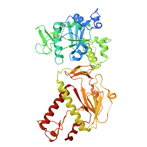
9NAZ, 9NFP, 9NHA, 9NHU, 9NIO, 9NJG - PubMed Abstract:
Point mutations in the exonuclease (ExoN) site of nonstructural protein 14 (NSP14) compromise the fitness of betacoronaviruses such as SARS-CoV-2, implicating NSP14 ExoN inhibition as an antiviral strategy. However, there are no advanced compounds that inhibit NSP14's ExoN activity. Building upon the reported crystal structures of two fragments bound to NSP14's ExoN site, we identified a series of 3,5-disubsituted pyrazoles that bound to and inhibited NSP14 ExoN. However, upon resynthesis, we discovered that these putative leads were false positives, perhaps due to contaminating divalent cations, which potently inhibit NSP14 ExoN. Our results provide a cautionary tale to the field about the sensitivity of NSP14 to divalent cations and illustrate the challenges associated with directly targeting the NSP14 ExoN site via fragment merging.
- Cleveland Clinic Center for Therapeutics Discovery, Cleveland Clinic Research, Cleveland, Ohio 44106, United States.
Organizational Affiliation: