9N8M | pdb_00009n8m

In situ sheathed flagellar FlaC filament in Vibrio cholerae.


Experimental Data Snapshot

  • Method: ELECTRON MICROSCOPY
  • Resolution: 3.16 Å
  • Aggregation State: CELL 
  • Reconstruction Method: SINGLE PARTICLE 

wwPDB Validation 3D Report Full Report

Validation slider image for 9N8M

This is version 1.2 of the entry. See complete history

Literature

Structures of the sheathed flagellum reveal mechanisms of assembly and rotation in Vibrio cholerae.

Guo, W.Zhang, S.Park, J.H.Stanton, V.Asp, M.Herrera, H.Tai, J.B.Yue, J.Wang, J.Guo, J.Kumar, R.Botting, J.M.Wu, S.Yan, J.Klose, K.E.Yildiz, F.H.Liu, J.

(2025) Nat Microbiol 10: 3305-3314

  • DOI: https://doi.org/10.1038/s41564-025-02161-x
  • Primary Citation Related Structures: 
    9N8A, 9N8B, 9N8G, 9N8H, 9N8M, 9P7R

  • PubMed Abstract: 

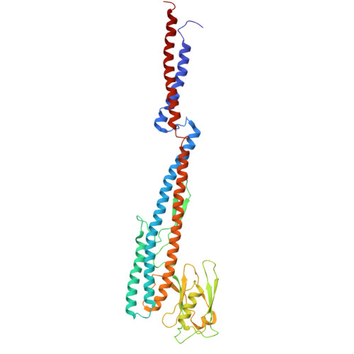
    Motility promotes the complex life cycle and infectious capabilities of Vibrio cholerae and is driven by rotation of a single polar flagellum. The flagellar filament comprises four flagellin proteins (FlaA-D) and is covered by a membranous sheath continuous with the outer membrane. Here we combine in situ cryo-electron microscopy single-particle analysis, fluorescence microscopy and molecular genetics to determine 2.92-3.43 Å structures of the sheathed flagellar filament from intact bacteria. Our data reveal the spatial arrangement of FlaA-D, showing that FlaA localizes at the cell pole and functions as a template for filament assembly involving multiple flagellins. Unlike unsheathed flagellar filaments, the sheathed filament from V. cholerae possesses a highly conserved core but a smooth, hydrophilic surface adjacent to the membranous sheath. A tiny conformational change at the single flagellin level results in a supercoiled filament and curved membranous sheath, supporting a model wherein the filament rotates separately from the sheath, enabling the distinct motility of V. cholerae.


  • Organizational Affiliation
    • Department of Microbial Pathogenesis, Yale School of Medicine, New Haven, CT, USA. wangbiao.guo@yale.edu.

Macromolecule Content 

  • Total Structure Weight: 1,318.16 kDa 
  • Atom Count: 92,202 
  • Modeled Residue Count: 12,441 
  • Deposited Residue Count: 12,441 
  • Unique protein chains: 1

Macromolecules

Find similar proteins by:|  3D Structure
Entity ID: 1
MoleculeChains  Sequence LengthOrganismDetailsImage
Flagellin C377Vibrio cholerae O1 biovar El Tor str. N16961Mutation(s): 0 
UniProt
Find proteins for P0C6C5 (Vibrio cholerae serotype O1 (strain ATCC 39315 / El Tor Inaba N16961))
Explore P0C6C5 
Go to UniProtKB:  P0C6C5
Entity Groups
Sequence Clusters30% Identity50% Identity70% Identity90% Identity95% Identity100% Identity
UniProt GroupP0C6C5
Sequence Annotations
Expand
Reference Sequence

Experimental Data & Validation

Experimental Data

  • Method: ELECTRON MICROSCOPY
  • Resolution: 3.16 Å
  • Aggregation State: CELL 
  • Reconstruction Method: SINGLE PARTICLE 
EM Software:
TaskSoftware PackageVersion
MODEL REFINEMENTPHENIX1.21.2_5419
RECONSTRUCTIONcryoSPARC

Structure Validation

View Full Validation Report


Entry History 

& Funding Information

Deposition Data


Funding OrganizationLocationGrant Number
National Institutes of Health/National Institute Of Allergy and Infectious Diseases (NIH/NIAID)United StatesR01AI087946
National Institutes of Health/National Institute Of Allergy and Infectious Diseases (NIH/NIAID)United StatesR01AI132818

Revision History  (Full details and data files)

  • Version 1.0: 2025-09-24
    Type: Initial release
  • Version 1.1: 2025-11-12
    Changes: Data collection, Database references
  • Version 1.2: 2025-12-10
    Changes: Data collection, Database references