9N5L | pdb_00009n5l

The Red Crowned-Crane Parvovirus Capsid


Experimental Data Snapshot

  • Method: ELECTRON MICROSCOPY
  • Resolution: 2.66 Å
  • Aggregation State: PARTICLE 
  • Reconstruction Method: SINGLE PARTICLE 

wwPDB Validation   3D Report Full Report


This is version 1.2 of the entry. See complete history


Literature

Birds of a feather flock together: structural characterization of red-crowned crane and turkey aveparvoviruses.

Hsi, J.Mietzsch, M.Patney, M.Chen, S.Sawh-Gopal, A.Chipman, P.McKenna, R.

(2025) J Virol 99: e0011025-e0011025

  • DOI: https://doi.org/10.1128/jvi.00110-25
  • Primary Citation of Related Structures:  
    9N5L, 9N5M

  • PubMed Abstract: 

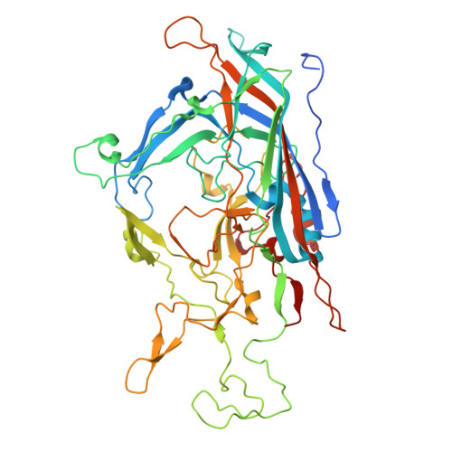
    The parvoviruses have non-enveloped T = 1 icosahedral capsids that are ~25 nm in diameter, which package linear single-stranded DNA and infect a wide range of hosts. To date, parvoviruses affecting birds have been identified in two genera: Ave - and Dependoparvovirus , and no capsid structures have been determined for any member of Aveparvovirus . This study investigates two bird viruses of this genus: red-crowned crane parvovirus (RCPV) and turkey parvovirus (TuPV). While the pathogenicity of RCPV is currently unknown, TuPV has been associated with gastrointestinal diseases, especially in juvenile birds. High-resolution structures of the RCPV and TuPV capsids were determined by cryo-electron microscopy at a resolution of 2.66 Å and 2.35 Å, respectively. A structural comparison of the RCPV and TuPV capsids shows that they exhibit many conserved features, such as a channel at the five-fold symmetry axis and surface depressions at the two-fold axis, as previously observed in parvoviruses from other genera. However, major structural differences were observed at the three-fold axes, with both RCPV and TuPV displaying recessed protrusions. In addition, terminal sialic acid was identified as a potential glycan receptor for RCPV. This study extends the architectural portfolio of structural parvovirology and will provide insights into parvoviral diversity and characterization of these viruses.IMPORTANCEThis study presents the capsid structures of two aveparvoviruses, red-crowned crane parvovirus (RCPV) and turkey parvovirus (TuPV), extending the structural repertoire of the Parvoviridae . While the pathogenicity of RCPV is unknown, the red-crowned cranes are among the rarest crane species. To date, very few virological studies have been conducted for this rare avian species, and understanding their virome could contribute to conservation efforts. Additionally, several studies have previously suggested that TuPV is associated with cases of enteric disease syndrome. To date, no commercial antivirals or vaccines are available for TuPV. The structural characterization of its capsid may contribute toward the development of a treatment to control the spread of infection.


  • Organizational Affiliation
    • Department of Biochemistry and Molecular Biology, Center for Structural Biology, McKnight Brain Institute, College of Medicine, University of Florida, Gainesville, Florida, USA.

Macromolecules
Find similar proteins by:  (by identity cutoff)  |  3D Structure
Entity ID: 1
MoleculeChains Sequence LengthOrganismDetailsImage
Major capsid protein531Red-crowned crane parvovirusMutation(s): 0 
Gene Names: VP2
UniProt
Find proteins for A0A2K9YN82 (Red-crowned crane parvovirus)
Explore A0A2K9YN82 
Go to UniProtKB:  A0A2K9YN82
Entity Groups  
Sequence Clusters30% Identity50% Identity70% Identity90% Identity95% Identity100% Identity
UniProt GroupA0A2K9YN82
Sequence Annotations
Expand
  • Reference Sequence
Small Molecules
Ligands 1 Unique
IDChains Name / Formula / InChI Key2D Diagram3D Interactions
MG
Query on MG

Download Ideal Coordinates CCD File 
AC [auth J]
AD [auth W]
AE [auth j]
AF [auth w]
BC [auth J]
AC [auth J],
AD [auth W],
AE [auth j],
AF [auth w],
BC [auth J],
BD [auth W],
BE [auth j],
BF [auth w],
CC [auth K],
CD [auth X],
CE [auth k],
CF [auth x],
DC [auth K],
DD [auth X],
DE [auth k],
DF [auth x],
EC [auth L],
ED [auth Y],
EE [auth l],
EF [auth y],
FC [auth L],
FD [auth Y],
FE [auth l],
FF [auth y],
GC [auth M],
GD [auth Z],
GE [auth m],
GF [auth z],
HC [auth M],
HD [auth Z],
HE [auth m],
HF [auth z],
IB [auth A],
IC [auth N],
ID [auth a],
IE [auth n],
IF [auth 1],
JB [auth A],
JC [auth N],
JD [auth a],
JE [auth n],
JF [auth 1],
KB [auth B],
KC [auth O],
KD [auth b],
KE [auth o],
KF [auth 2],
LB [auth B],
LC [auth O],
LD [auth b],
LE [auth o],
LF [auth 2],
MB [auth C],
MC [auth P],
MD [auth c],
ME [auth p],
MF [auth 3],
NB [auth C],
NC [auth P],
ND [auth c],
NE [auth p],
NF [auth 3],
OB [auth D],
OC [auth Q],
OD [auth d],
OE [auth q],
OF [auth 4],
PB [auth D],
PC [auth Q],
PD [auth d],
PE [auth q],
PF [auth 4],
QB [auth E],
QC [auth R],
QD [auth e],
QE [auth r],
QF [auth 5],
RB [auth E],
RC [auth R],
RD [auth e],
RE [auth r],
RF [auth 5],
SB [auth F],
SC [auth S],
SD [auth f],
SE [auth s],
SF [auth 6],
TB [auth F],
TC [auth S],
TD [auth f],
TE [auth s],
TF [auth 6],
UB [auth G],
UC [auth T],
UD [auth g],
UE [auth t],
UF [auth 7],
VB [auth G],
VC [auth T],
VD [auth g],
VE [auth t],
VF [auth 7],
WB [auth H],
WC [auth U],
WD [auth h],
WE [auth u],
WF [auth 8],
XB [auth H],
XC [auth U],
XD [auth h],
XE [auth u],
XF [auth 8],
YB [auth I],
YC [auth V],
YD [auth i],
YE [auth v],
ZB [auth I],
ZC [auth V],
ZD [auth i],
ZE [auth v]
MAGNESIUM ION
Mg
JLVVSXFLKOJNIY-UHFFFAOYSA-N
Experimental Data & Validation

Experimental Data

  • Method: ELECTRON MICROSCOPY
  • Resolution: 2.66 Å
  • Aggregation State: PARTICLE 
  • Reconstruction Method: SINGLE PARTICLE 
EM Software:
TaskSoftware PackageVersion
MODEL REFINEMENTPHENIX1.10-2155_2155

Structure Validation

View Full Validation Report


Entry History & Funding Information

Deposition Data


Funding OrganizationLocationGrant Number
National Science Foundation (NSF, United States)United StatesDGE-2236414
National Institutes of Health/National Institute of General Medical Sciences (NIH/NIGMS)United StatesGM082946

Revision History  (Full details and data files)

  • Version 1.0: 2025-07-02
    Type: Initial release
  • Version 1.1: 2025-07-16
    Changes: Data collection, Database references
  • Version 1.2: 2025-07-30
    Changes: Data collection, Database references