9N2G | pdb_00009n2g

Structure of apo Mtb NapA(16-90)


Experimental Data Snapshot

  • Method: X-RAY DIFFRACTION
  • Resolution: 2.39 Å
  • R-Value Free: 
    0.243 (Depositor), 0.244 (DCC) 
  • R-Value Work: 
    0.227 (Depositor), 0.228 (DCC) 
  • R-Value Observed: 
    0.230 (Depositor) 

Starting Model: experimental
View more details

wwPDB Validation 3D Report Full Report

Validation slider image for 9N2G

This is version 1.1 of the entry. See complete history

Literature

Structural Studies on the M. tuberculosis Nucleoid-associated-Protein, NapA, Indicates DNA Bridging Mechanism.

Schumacher, M.A.Singh, R.R.Salinas, R.

(2025) J Mol Biology 437: 169486-169486

  • DOI: https://doi.org/10.1016/j.jmb.2025.169486
  • Primary Citation Related Structures: 
    9N2G, 9N2N

  • PubMed Abstract: 

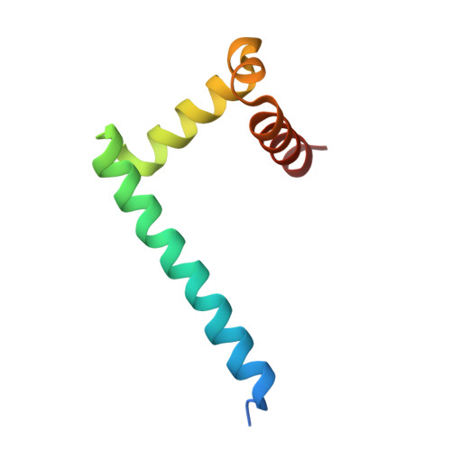
    Nucleoid-associated proteins (NAPs) play central roles in bacterial chromosome organization and DNA processes. Interestingly, Mycobacterium tuberculosis (Mtb) lacks most common NAPs and only recently have NAPs been uncovered in this bacterium. One such protein, NapA, was revealed to be an essential Mtb NAP that can bridge DNA. NapA shows no sequence homology to any protein and hence its DNA-binding functions remain unclear. Here we describe structures of apo NapA and a DNA-bound complex of NapA. The NapA structures reveal a dimeric fold for the protein, which is supported by mass photometry analyses, with each subunit comprised of an extended α1 helix and C-terminal three-helix module. The α1 helices combine to form a helical-bundle dimer scaffold that forms dimer-of-dimers at elevated protein concentrations. Each NapA dimer projects two DNA interacting elements, that bind and link between DNA sites. Combined these studies provide mechanistic insight into the DNA binding and bridging capabilities of a unique NAP that appears broadly conserved among most Actinobacteria.


  • Organizational Affiliation
    • Department of Biochemistry, 307 Research Dr., Box 3711, Duke University Medical Center, Durham, NC 27710, USA. Electronic address: maria.schumacher@duke.edu.

Macromolecule Content 

  • Total Structure Weight: 18.24 kDa 
  • Atom Count: 1,184 
  • Modeled Residue Count: 143 
  • Deposited Residue Count: 156 
  • Unique protein chains: 1

Macromolecules

Find similar proteins by:|  3D Structure
Entity ID: 1
MoleculeChains  Sequence LengthOrganismDetailsImage
Fis family transcriptional regulatorA [auth U],
B [auth A]
78Mycobacterium tuberculosisMutation(s): 0 
Gene Names: 
UniProt
Find proteins for P96276 (Mycobacterium tuberculosis (strain ATCC 25618 / H37Rv))
Explore P96276 
Go to UniProtKB:  P96276
Entity Groups
Sequence Clusters30% Identity50% Identity70% Identity90% Identity95% Identity100% Identity
UniProt GroupP96276
Sequence Annotations
Expand
Reference Sequence

Experimental Data & Validation

Experimental Data

  • Method: X-RAY DIFFRACTION
  • Resolution: 2.39 Å
  • R-Value Free:  0.243 (Depositor), 0.244 (DCC) 
  • R-Value Work:  0.227 (Depositor), 0.228 (DCC) 
  • R-Value Observed: 0.230 (Depositor) 
Space Group: I 1 2 1
Unit Cell:
Length ( Å )Angle ( ˚ )
a = 41.9α = 90
b = 50.359β = 104.17
c = 62.948γ = 90
Software Package:
Software NamePurpose
PHENIXrefinement
iMOSFLMdata reduction
Aimlessdata scaling
Cootmodel building
MOLREPphasing

Structure Validation

View Full Validation Report


Entry History 

& Funding Information

Deposition Data


Funding OrganizationLocationGrant Number
National Institutes of Health/National Institute of General Medical Sciences (NIH/NIGMS)United StatesR35GM130290

Revision History  (Full details and data files)

  • Version 1.0: 2025-10-22
    Type: Initial release
  • Version 1.1: 2026-05-06
    Changes: Database references