The Heme Oxygenase-Like Diiron Enzyme HrmI Reveals Altered Regulatory Mechanisms for Dioxygen Activation and Substrate N-Oxygenation.
Skirboll, S.S., Gangopadhyay, M., Phan, H.N., Hartsell, J., Mudireddy, A., Hilovsky, D., Swartz, P.D., Liu, X., Guo, Y., Makris, T.M.(2025) J Am Chem Soc 147: 30210-30221
- PubMed: 40774922 Search on PubMed
- DOI: https://doi.org/10.1021/jacs.5c08814
- Primary Citation Related Structures:
9N1A, 9N1E, 9N1X, 9N2A, 9NH9 - PubMed Abstract:
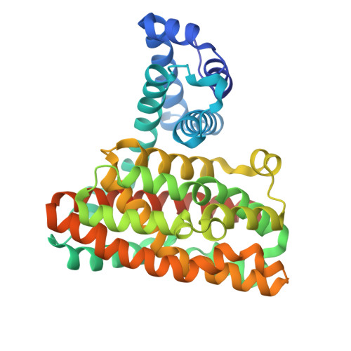
Nonheme diiron enzymes activate dioxygen (O 2 ) to affect various biochemical outcomes. HrmI, a member of the recently discovered and functionally versatile heme oxygenase-like dimetal oxidase/oxygenase (HDO) superfamily, catalyzes the N-oxygenation of L-Lysine to yield 6-nitronorleucine for the biosynthesis of the antibiotic hormaomycin. Unlike other characterized HDO N-oxygenases that have an additional carboxylate ligand thought to be key for regulating dioxygen activation and ensuing N-oxygenation, the predicted primary coordination sphere of HrmI resembles those of HDOs that instead perform C-C fragmentation of substrates. We show that diferrous HrmI reacts with O 2 in a substrate-independent manner to form a presumptive μ-1,2 (Fe 3+ ) 2 peroxo (or P ) intermediate common to the catalytic scheme of many HDOs. P is rapidly converted to a second species with both optical and Mössbauer properties that resemble an activated peroxodiferric adduct ( P' ). The substrate-dependent acceleration of P' decay suggests that it, rather than P , initiates l-Lysine metabolism. X-ray crystallographic studies of HrmI in several redox and ligand-bound states provide a stepwise view of structural changes during catalysis and, together with analytical approaches, capture a hydroxylamino metabolic intermediate en route to 6-nitronorleucine formation. The activation of peroxo species provides a key strategy that enables functional adaptation within the widely distributed HDO structural scaffold.
- Department of Molecular and Structural Biochemistry, North Carolina State University, Raleigh, North Carolina 27695, United States.
Organizational Affiliation: