9MQY | pdb_00009mqy

Structure of mycobacterial NDH2 (type II NADH:quinone oxidoreductase)


Experimental Data Snapshot

  • Method: ELECTRON MICROSCOPY
  • Resolution: 3.00 Å
  • Aggregation State: PARTICLE 
  • Reconstruction Method: SINGLE PARTICLE 

wwPDB Validation   3D Report Full Report


This is version 1.3 of the entry. See complete history


Literature

Structure of Mycobacterial NDH-2 Bound to a 2-Mercapto-Quinazolinone Inhibitor.

Liang, Y.Bueler, S.A.Cook, G.M.Rubinstein, J.L.

(2025) J Med Chem 68: 7579-7591

  • DOI: https://doi.org/10.1021/acs.jmedchem.5c00049
  • Primary Citation of Related Structures:  
    9MQY, 9MQZ

  • PubMed Abstract: 

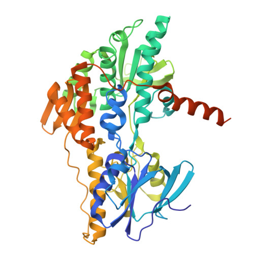
    Mycobacterial type II NADH dehydrogenase (NDH-2) is a promising drug target because of its central role in energy metabolism in Mycobacterium tuberculosis and other pathogens, and because it lacks a known mammalian homologue. To facilitate optimization of lead compounds, we used electron cryomicroscopy (cryo-EM) to determine the structure of NDH-2 from Mycobacterium smegmatis , a fast-growing nonpathogenic model for respiration in M. tuberculosis . The structure shows that active mycobacterial NDH-2 is dimeric, with an arrangement of monomers in the dimer that differs from the arrangement described for other prokaryotic NDH-2 dimers, instead resembling dimers formed by NDH-2 in the eukaryotes Saccharomyces cerevisiae and Plasmodium falciparum . A structure of the enzyme bound to a 2-mercapto-quinazolinone inhibitor shows that the compound interacts directly with the flavin adenine dinucleotide cofactor, blocking the menaquinone-reducing site. These results reveal structural elements of NDH-2 that could be used to design specific inhibitors of the mycobacterial enzyme.


  • Organizational Affiliation
    • Molecular Medicine Program, The Hospital for Sick Children, Toronto, ON M5G 0A4, Canada.

Macromolecules
Find similar proteins by:  (by identity cutoff)  |  3D Structure
Entity ID: 1
MoleculeChains Sequence LengthOrganismDetailsImage
NADH:ubiquinone reductase (non-electrogenic)
A, B
494Mycolicibacterium smegmatis MC2 155Mutation(s): 0 
EC: 1.6.5.9
UniProt
Find proteins for P95160 (Mycobacterium tuberculosis (strain ATCC 25618 / H37Rv))
Explore P95160 
Go to UniProtKB:  P95160
Entity Groups  
Sequence Clusters30% Identity50% Identity70% Identity90% Identity95% Identity100% Identity
UniProt GroupP95160
Sequence Annotations
Expand
  • Reference Sequence
Experimental Data & Validation

Experimental Data

  • Method: ELECTRON MICROSCOPY
  • Resolution: 3.00 Å
  • Aggregation State: PARTICLE 
  • Reconstruction Method: SINGLE PARTICLE 
EM Software:
TaskSoftware PackageVersion
RECONSTRUCTIONcryoSPARC4.6.2
MODEL REFINEMENTPHENIX

Structure Validation

View Full Validation Report


Entry History & Funding Information

Deposition Data


Funding OrganizationLocationGrant Number
Canadian Institutes of Health Research (CIHR)CanadaPJT191893

Revision History  (Full details and data files)

  • Version 1.0: 2025-01-22
    Type: Initial release
  • Version 1.1: 2025-04-02
    Changes: Data collection, Database references
  • Version 1.2: 2025-04-23
    Changes: Data collection, Database references
  • Version 1.3: 2025-05-21
    Changes: Data collection