The Position of Indole Methylation Controls the Structure, DNA Binding, and Cellular Functions of Mithramycin SA-Trp Analogues.
Hou, C., Bhosale, S., Yasuda, K., Yetirajam, R., Leggas, M., Rohr, J., Tsodikov, O.V.(2025) Chembiochem 26: e202401084-e202401084
- PubMed: 40246689 Search on PubMed
- DOI: https://doi.org/10.1002/cbic.202401084
- Primary Citation Related Structures:
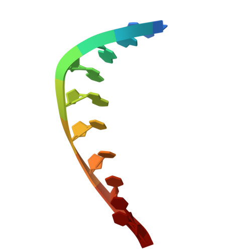
9MOZ, 9MP1 - PubMed Abstract:
Mithramycin (MTM) is a polyketide anti-cancer natural product, which functions by noncovalent binding to DNA in the minor groove without intercalation, resulting in inhibiting transcription at G/C-rich promoters. MTM is a potent inhibitor of cancer cells, such as Ewing sarcoma, driven by abnormal fusions involving ETS family transcription factors FLI1 and ERG. However, MTM is rather toxic and non-selective; therefore, safer, selective analogues of MTM are required for use in clinic as anti-cancer drugs. In this study by using a combination of X-ray crystallographic, biophysical, cell and molecular biological techniques, we explored structural and functional consequences of 3-side chain methylation at positions 5, 6 and 7 of the indole ring of the potent analogue MTM SA-Trp. We showed that the conformation of the analogues in complexes with DNA, their DNA binding function, cytotoxicity, selectivity and potency as transcription antagonists depended on the position of the methylation. MTM SA-5-methyl-Trp emerged as the most selective analogue, presumably due to the right balance of the DNA binding and the solvent exposure of the 3-side chain. This study demonstrates that minor chemical changes can have strong effects in analogue development and paves a way to further development of next generation MTM analogues.
- University of Kentucky, Pharmaceutical Sciences, Lee T. Todd, Jr Building, 789 South Limestone St., Room 425, 40536, Lexington, UNITED STATES OF AMERICA.
Organizational Affiliation: