Exploring the Structural Divergence of HIV and SRLV Lentiviral Capsids.
Arizaga Jr., F., Freniere, C., Rey, J.S., Cook, M., Wu, C., Perilla, J.R., Xiong, Y.(2025) J Am Chem Soc 147: 32883-32895
- PubMed: 40878534 Search on PubMed
- DOI: https://doi.org/10.1021/jacs.5c09436
- Primary Citation Related Structures:
9MKP, 9MKQ, 9MKR, 9MKS - PubMed Abstract:
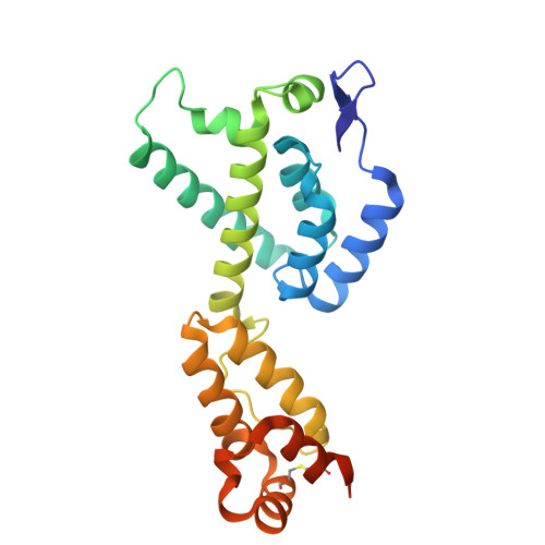
Lentiviruses require a mature capsid to package and traffic their viral genome for successful infection and propagation. Although the HIV-1 capsid structure has been extensively studied, structural information is lacking for other lentiviral capsids, limiting our understanding. Using cryo-electron microscopy (cryo-EM) and a liposome-templating system, we assembled capsid-like particles (CLPs) and resolved capsid protein (CA) pentamer and hexamer lattice structures from the two major phylogenetic groups of small ruminant lentiviruses (SRLVs). These structures exhibit an overall lattice organization like HIV-1 but differ in key characteristics, notably the absence of inositol hexakisphosphate (IP6) in the SRLV CA lattice─a critical factor for HIV-1 capsid assembly and function. Additionally, SRLV CA pentamers show a unique N-terminal domain orientation, providing insights into SRLV capsid assembly mechanisms. These observations, together with our molecular dynamics (MD) simulation, results suggest a possible mechanism for importing deoxynucleotide triphosphate (dNTP) molecules into SRLV capsids. Furthermore, key regions of host factor interaction, such as the CypA binding motifs, have diverged in the SRLV CA assemblies. Our results contribute to understanding the SRLV lentiviral capsids which may facilitate structure-based inhibitor design strategies.
- Department of Molecular Biophysics and Biochemistry, Yale University, New Haven, Connecticut 06511, United States.
Organizational Affiliation: