Mechanistic and evolutionary insights into a family of aminoacyl-tRNA deacylases that protects against canavanine toxicity.
Maldonado, J.S., Sepulveda, S., Karthikeyan, S., Shirakawa, K.T., Merced, I., Radecki, A.A., Douglas, J., Peti, W., Page, R., Vargas-Rodriguez, O.(2025) Nucleic Acids Res 53
- PubMed: 40973455 Search on PubMedSearch on PubMed Central
- DOI: https://doi.org/10.1093/nar/gkaf922
- Primary Citation Related Structures:
9MKL - PubMed Abstract:
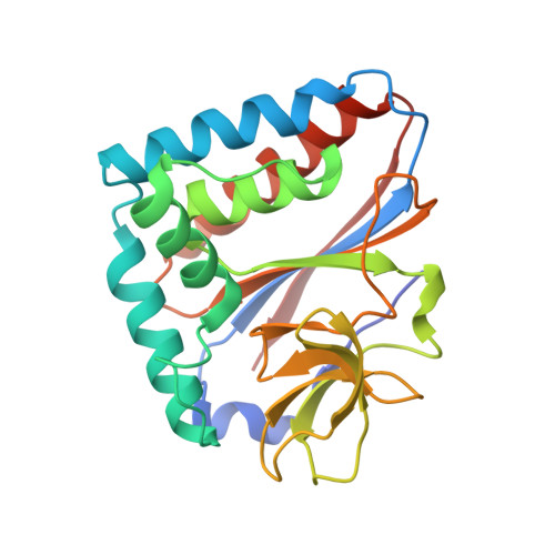
Aminoacyl-tRNA deacylases safeguard the accurate translation of the genetic code by hydrolyzing incorrectly synthesized aminoacyl-tRNAs. Canavanyl-tRNA deacylase (CtdA) was recently shown to protect cells against the toxicity of canavanine (Can), a nonproteinogenic amino acid synthesized and accumulated by leguminous plants. In most organisms, Can is ligated to tRNAArg, causing translation of arginine codons with Can. CtdA prevents Can toxicity by hydrolyzing canavanyl-tRNAArg. Here, we investigated the function, structure, substrate specificity, phylogenetic distribution, and evolution of CtdA. We show that CtdA is essential for preventing Can cytotoxicity in Salmonella enterica, and its heterologous expression can also protect Escherichia coli. By determining the structure of CtdA, we identified its putative binding pocket and residues that modulate enzymatic activity and specificity. We also found that CtdA displays robust specificity for the canavanyl moiety, a feature that contributes to maintaining arginyl-tRNAArg levels unaffected. Finally, we showed that despite their structural homology, CtdA and the aminoacyl-tRNA hydrolytic domain of phenylalanyl-tRNA synthetase are functionally and evolutionarily divergent. Collectively, these results substantially expand our understanding of the CtdA family, providing new insights into its structure, function, and evolution. This work also highlights the diverse mechanisms, unique to each organism, that ensure faithful translation of the genetic code.
- Department of Molecular Biology and Biophysics, University of Connecticut School of Medicine, Farmington, CT 06030, United States.
Organizational Affiliation: