9MIV | pdb_00009miv

Native tagless Lassa virus spike complex pH 6.0


Experimental Data Snapshot

  • Method: ELECTRON MICROSCOPY
  • Resolution: 2.02 Å
  • Aggregation State: PARTICLE 
  • Reconstruction Method: SINGLE PARTICLE 

wwPDB Validation   3D Report Full Report


This is version 1.2 of the entry. See complete history


Literature

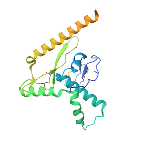
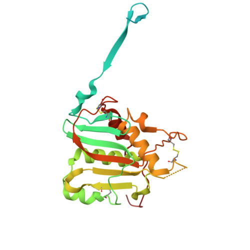
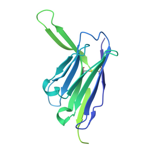
pH-induced conformational changes and inhibition of the Lassa virus spike complex.

Katz, M.Cohen-Dvashi, H.Borni, S.Ruedas, J.Henkel, G.McCormack, K.Diskin, R.

(2025) Cell Host Microbe 33: 1577-1588.e7

  • DOI: https://doi.org/10.1016/j.chom.2025.07.020
  • Primary Citation of Related Structures:  
    9MHE, 9MIV, 9MIY, 9MJ1, 9MJ2, 9R8U

  • PubMed Abstract: 

    Lassa virus (LASV) is a devastating human pathogen with no vaccines and limited therapeutics. The LASV class-I spike complex engages target cells via binding its primary host receptor, matriglycan, followed by macropinocytosis and binding of its secondary receptor, lysosomal-associated membrane protein 1 (LAMP1), to trigger virus fusion. This process occurs across multiple pH-dependent steps, but the molecular events remain largely unknown. Through high-resolution structures, we study the pH-induced conformational changes of the spike preceding membrane fusion. We reveal pH-sensitive metal coordination sites that control the integrity of the spike's native state, elucidate a reorganization of the spike's transmembrane region, and provide a mechanism for dissociation from its primary receptor. Using the entry inhibitor ARN-75039, we validate our findings and establish the molecular basis for the binding and function of this investigational drug. These data define the molecular basis for the cell entry of LASV and will promote efforts in combating this virus and potentially related viral pathogens.


  • Organizational Affiliation
    • Department of Chemical and Structural Biology, Weizmann Institute of Science, 7610001 Rehovot, Israel.

Macromolecules
Find similar proteins by:  (by identity cutoff)  |  3D Structure
Entity ID: 1
MoleculeChains Sequence LengthOrganismDetailsImage
Glycoprotein G2A [auth a],
K [auth b],
L [auth c]
232Lassa virus JosiahMutation(s): 0 
Gene Names: GPCGP-C
UniProt
Find proteins for P08669 (Lassa virus (strain Mouse/Sierra Leone/Josiah/1976))
Explore P08669 
Go to UniProtKB:  P08669
Entity Groups  
Sequence Clusters30% Identity50% Identity70% Identity90% Identity95% Identity100% Identity
UniProt GroupP08669
Glycosylation
Glycosylation Sites: 3
Sequence Annotations
Expand
  • Reference Sequence
Find similar proteins by:  (by identity cutoff)  |  3D Structure
Entity ID: 2
MoleculeChains Sequence LengthOrganismDetailsImage
Glycoprotein G1B [auth A],
C [auth B],
D [auth C]
259Lassa virus JosiahMutation(s): 0 
Gene Names: GPCGP-C
UniProt
Find proteins for P08669 (Lassa virus (strain Mouse/Sierra Leone/Josiah/1976))
Explore P08669 
Go to UniProtKB:  P08669
Entity Groups  
Sequence Clusters30% Identity50% Identity70% Identity90% Identity95% Identity100% Identity
UniProt GroupP08669
Glycosylation
Glycosylation Sites: 7
Sequence Annotations
Expand
  • Reference Sequence
Find similar proteins by:  (by identity cutoff)  |  3D Structure
Entity ID: 3
MoleculeChains Sequence LengthOrganismDetailsImage
12.1F Heavy chainE [auth H],
I [auth D],
J [auth F]
249Homo sapiensMutation(s): 0 
Entity Groups  
Sequence Clusters30% Identity50% Identity70% Identity90% Identity95% Identity100% Identity
Sequence Annotations
Expand
  • Reference Sequence
Find similar proteins by:  (by identity cutoff)  |  3D Structure
Entity ID: 4
MoleculeChains Sequence LengthOrganismDetailsImage
12.1F Light chainF [auth L],
G [auth E],
H [auth G]
214Homo sapiensMutation(s): 0 
Entity Groups  
Sequence Clusters30% Identity50% Identity70% Identity90% Identity95% Identity100% Identity
Sequence Annotations
Expand
  • Reference Sequence
Oligosaccharides

Help

Entity ID: 5
MoleculeChains Length2D Diagram Glycosylation3D Interactions
2-acetamido-2-deoxy-beta-D-glucopyranose-(1-2)-alpha-D-mannopyranose-(1-3)-[alpha-D-mannopyranose-(1-3)-alpha-D-mannopyranose-(1-6)]beta-D-mannopyranose-(1-4)-2-acetamido-2-deoxy-beta-D-glucopyranose-(1-4)-2-acetamido-2-deoxy-beta-D-glucopyranoseFA [auth f],
GA [auth g],
M [auth I]
7N-Glycosylation
Glycosylation Resources
GlyTouCan:  G53168IY
GlyCosmos:  G53168IY
GlyGen:  G53168IY
Entity ID: 6
MoleculeChains Length2D Diagram Glycosylation3D Interactions
beta-D-mannopyranose-(1-4)-2-acetamido-2-deoxy-beta-D-glucopyranose-(1-4)-2-acetamido-2-deoxy-beta-D-glucopyranoseN [auth J],
P [auth M],
U [auth R],
Z [auth W]
3N-Glycosylation
Glycosylation Resources
GlyTouCan:  G15407YE
GlyCosmos:  G15407YE
GlyGen:  G15407YE
Entity ID: 7
MoleculeChains Length2D Diagram Glycosylation3D Interactions
alpha-D-mannopyranose-(1-6)-beta-D-mannopyranose-(1-4)-2-acetamido-2-deoxy-beta-D-glucopyranose-(1-4)-[alpha-L-fucopyranose-(1-6)]2-acetamido-2-deoxy-beta-D-glucopyranoseO [auth K],
T [auth Q],
Y [auth V]
5N-Glycosylation
Glycosylation Resources
GlyTouCan:  G00395TQ
GlyCosmos:  G00395TQ
GlyGen:  G00395TQ
Entity ID: 8
MoleculeChains Length2D Diagram Glycosylation3D Interactions
beta-D-galactopyranose-(1-4)-2-acetamido-2-deoxy-beta-D-glucopyranose-(1-2)-alpha-D-mannopyranose-(1-3)-[alpha-D-mannopyranose-(1-6)]beta-D-mannopyranose-(1-4)-2-acetamido-2-deoxy-beta-D-glucopyranose-(1-4)-2-acetamido-2-deoxy-beta-D-glucopyranoseAA [auth X],
Q [auth N],
V [auth S]
7N-Glycosylation
Glycosylation Resources
GlyTouCan:  G69411IG
GlyCosmos:  G69411IG
GlyGen:  G69411IG
Entity ID: 9
MoleculeChains Length2D Diagram Glycosylation3D Interactions
alpha-D-mannopyranose-(1-6)-alpha-D-mannopyranose-(1-3)-beta-D-mannopyranose-(1-4)-2-acetamido-2-deoxy-beta-D-glucopyranose-(1-4)-[alpha-L-fucopyranose-(1-6)]2-acetamido-2-deoxy-beta-D-glucopyranoseR [auth O]6N-Glycosylation
Entity ID: 10
MoleculeChains Length2D Diagram Glycosylation3D Interactions
alpha-D-mannopyranose-(1-3)-beta-D-mannopyranose-(1-4)-2-acetamido-2-deoxy-beta-D-glucopyranose-(1-4)-2-acetamido-2-deoxy-beta-D-glucopyranoseS [auth P],
X [auth U]
4N-Glycosylation
Glycosylation Resources
GlyTouCan:  G81315DD
GlyCosmos:  G81315DD
GlyGen:  G81315DD
Entity ID: 11
MoleculeChains Length2D Diagram Glycosylation3D Interactions
alpha-D-mannopyranose-(1-3)-beta-D-mannopyranose-(1-4)-2-acetamido-2-deoxy-beta-D-glucopyranose-(1-4)-[alpha-L-fucopyranose-(1-6)]2-acetamido-2-deoxy-beta-D-glucopyranoseBA [auth Y],
W [auth T]
5N-Glycosylation
Glycosylation Resources
GlyTouCan:  G42466VF
GlyCosmos:  G42466VF
GlyGen:  G42466VF
Entity ID: 12
MoleculeChains Length2D Diagram Glycosylation3D Interactions
alpha-D-xylopyranose-(1-3)-beta-D-glucopyranuronic acid-(1-3)-alpha-D-xylopyranoseCA [auth Z],
DA [auth d],
EA [auth e]
3N/A
Glycosylation Resources
GlyTouCan:  G54924MX
GlyCosmos:  G54924MX
Small Molecules
Ligands 2 Unique
IDChains Name / Formula / InChI Key2D Diagram3D Interactions
NAG
Query on NAG

Download Ideal Coordinates CCD File 
HA [auth a]
IA [auth a]
MA [auth A]
NA [auth A]
OA [auth B]
HA [auth a],
IA [auth a],
MA [auth A],
NA [auth A],
OA [auth B],
PA [auth B],
QA [auth C],
RA [auth C],
SA [auth b],
TA [auth b],
UA [auth c],
VA [auth c]
2-acetamido-2-deoxy-beta-D-glucopyranose
C8 H15 N O6
OVRNDRQMDRJTHS-FMDGEEDCSA-N
UNX
Query on UNX

Download Ideal Coordinates CCD File 
JA [auth a],
KA [auth a],
LA [auth a]
UNKNOWN ATOM OR ION
X
Experimental Data & Validation

Experimental Data

  • Method: ELECTRON MICROSCOPY
  • Resolution: 2.02 Å
  • Aggregation State: PARTICLE 
  • Reconstruction Method: SINGLE PARTICLE 
EM Software:
TaskSoftware PackageVersion
MODEL REFINEMENTPHENIXdev_5278

Structure Validation

View Full Validation Report


Entry History & Funding Information

Deposition Data


Funding OrganizationLocationGrant Number
Not funded--

Revision History  (Full details and data files)

  • Version 1.0: 2025-08-27
    Type: Initial release
  • Version 1.1: 2025-09-17
    Changes: Data collection, Database references
  • Version 1.2: 2025-09-24
    Changes: Data collection, Database references