9MIL | pdb_00009mil

Superoxide dismutase residues 28-39 with G37R mutation


Experimental Data Snapshot

  • Method: X-RAY DIFFRACTION
  • Resolution: 2.50 Å
  • R-Value Free: 
    0.263 (Depositor), 0.263 (DCC) 
  • R-Value Work: 
    0.219 (Depositor), 0.218 (DCC) 
  • R-Value Observed: 
    0.223 (Depositor) 

Starting Model: experimental
View more details

wwPDB Validation 3D Report Full Report

Validation slider image for 9MIL

This is version 1.1 of the entry. See complete history

Literature

Amyloid Oligomers: Expediting Crystal Growth and Revisiting the Corkscrew Structures.

Hirschbeck, S.S.Sawaya, M.R.Lindberg, E.T.Limbach, M.N.Jang, J.H.Lazar Cantrell, K.L.Eisenberg, D.S.Do, T.D.

(2025) J Am Chem Soc 147: 18594-18605

  • DOI: https://doi.org/10.1021/jacs.5c00656
  • Primary Citation Related Structures: 
    9MIL

  • PubMed Abstract: 

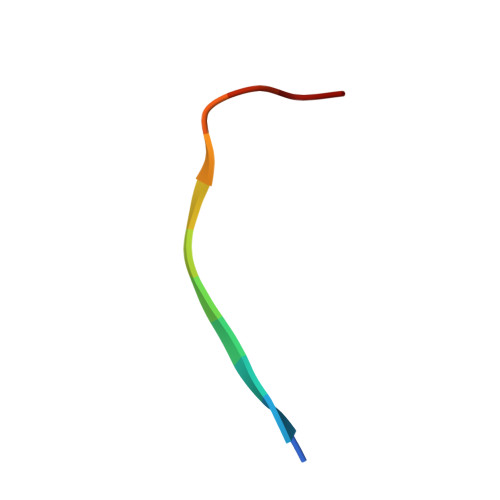
    Crystallizing soluble amyloid oligomers (AOs) presents a major challenge in studying disease-related mutations associated with amyloid diseases. The G37R mutation in superoxide dismutase 1 (SOD1) is linked to early onset amyotrophic lateral sclerosis (ALS), yet its toxic mechanism remains unclear. The transient nature and low solubility of AOs often complicate the production of high-quality crystals required for X-ray crystallography (XRC) analysis. To address these challenges, we employ native ion mobility spectrometry-mass spectrometry (IMS-MS) to screen SOD1 peptides and examine correlations between structural features that reflect AO stability, their sequence length, and specific mutations. In particular, previous studies showed that the P28K mutation in SOD1(28-38) enhances solubility, thus allowing the capture of AO corkscrew structures for both SOD1(28-38) P28K and SOD1(28-38) P28K, G37R . Building on these findings, we expanded our screening to include SOD1 peptides with longer sequences, identifying structural features in IMS-MS spectra that correlate with improved crystallization potential. This approach enabled us to distinguish the stabilizing effects of G37R from those of P28K, culminating in the successful determination of the first crystal structure of the SOD1 corkscrew containing the native proline.


  • Organizational Affiliation
    • Department of Chemistry, University of Tennessee, Knoxville, Tennessee 37996, United States.

Macromolecule Content 

  • Total Structure Weight: 14.84 kDa 
  • Atom Count: 1,049 
  • Modeled Residue Count: 120 
  • Deposited Residue Count: 120 
  • Unique protein chains: 1

Macromolecules

Find similar proteins by:  Sequence   |   3D Structure  
Entity ID: 1
MoleculeChains  Sequence LengthOrganismDetailsImage
Superoxide dismutase [Cu-Zn]
A, B, C, D, E
A, B, C, D, E, F, G, H, I, J
12Homo sapiensMutation(s): 1 
EC: 1.15.1.1 (PDB Primary Data), 1.8 (UniProt)
UniProt & NIH Common Fund Data Resources
Find proteins for P00441 (Homo sapiens)
Explore P00441 
Go to UniProtKB:  P00441
PHAROS:  P00441
GTEx:  ENSG00000142168 
Entity Groups
Sequence Clusters30% Identity50% Identity70% Identity90% Identity95% Identity100% Identity
UniProt GroupP00441
Sequence Annotations
Expand
Reference Sequence

Small Molecules

Ligands 3 Unique
IDChains Name / Formula / InChI Key2D Diagram3D Interactions
PG4

Query on PG4


Download:Ideal Coordinates CCD File
L [auth A],
R [auth J]
TETRAETHYLENE GLYCOL
C8 H18 O5
UWHCKJMYHZGTIT-UHFFFAOYSA-N
PEG

Query on PEG


Download:Ideal Coordinates CCD File
M [auth D]DI(HYDROXYETHYL)ETHER
C4 H10 O3
MTHSVFCYNBDYFN-UHFFFAOYSA-N
SO4

Query on SO4


Download:Ideal Coordinates CCD File
K [auth A],
N [auth F],
O [auth G],
P [auth H],
Q [auth I]
SULFATE ION
O4 S
QAOWNCQODCNURD-UHFFFAOYSA-L

Experimental Data & Validation

Experimental Data

  • Method: X-RAY DIFFRACTION
  • Resolution: 2.50 Å
  • R-Value Free:  0.263 (Depositor), 0.263 (DCC) 
  • R-Value Work:  0.219 (Depositor), 0.218 (DCC) 
  • R-Value Observed: 0.223 (Depositor) 
Space Group: P 31 2 1
Unit Cell:
Length ( Å )Angle ( ˚ )
a = 59.89α = 90
b = 59.89β = 90
c = 93.76γ = 120
Software Package:
Software NamePurpose
PHENIXrefinement
XSCALEdata scaling
XDSdata reduction
PHASERphasing

Structure Validation

View Full Validation Report


Entry History 

& Funding Information

Deposition Data


Funding OrganizationLocationGrant Number
National Institutes of Health/National Institute on Aging (NIH/NIA)United States1R01AG070895-01A1

Revision History  (Full details and data files)

  • Version 1.0: 2025-06-04
    Type: Initial release
  • Version 1.1: 2025-06-18
    Changes: Database references