


Crystal Structure of Human P38 alpha MAPK In Complex with MW01-14-064SRM
Minasov, G., Tokars, V.L., Roy, S.M., Watterson, D.M., Shuvalova, L.To be published.
Experimental Data Snapshot
Starting Model: experimental
View more details
Entity ID: 1 | |||||
|---|---|---|---|---|---|
| Molecule | Chains | Sequence Length | Organism | Details | Image |
| Mitogen-activated protein kinase 14 | 362 | Homo sapiens | Mutation(s): 0 Gene Names: MAPK14, CSBP, CSBP1, CSBP2, CSPB1, MXI2, SAPK2A EC: 2.7.11.24 | 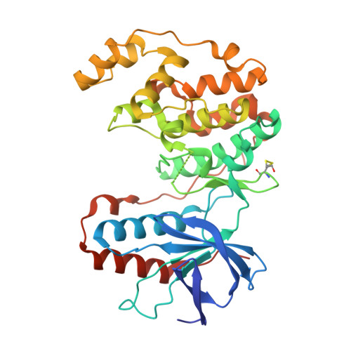 | |
UniProt & NIH Common Fund Data Resources | |||||
PHAROS: Q16539 GTEx: ENSG00000112062 | |||||
Entity Groups | |||||
| Sequence Clusters | 30% Identity50% Identity70% Identity90% Identity95% Identity100% Identity | ||||
| UniProt Group | Q16539 | ||||
Sequence AnnotationsExpand | |||||
Reference Sequence | |||||
| Ligands 5 Unique | |||||
|---|---|---|---|---|---|
| ID | Chains | Name / Formula / InChI Key | 2D Diagram | 3D Interactions | |
| A1BLA (Subject of Investigation/LOI) Download:Ideal Coordinates CCD File | B [auth A] | (5P)-N,N-dimethyl-5-(naphthalen-1-yl)-6-(pyridin-4-yl)pyrazin-2-amine C21 H18 N4 CDYRUKWQQPYHTN-UHFFFAOYSA-N |  | ||
| GG5 (Subject of Investigation/LOI) Download:Ideal Coordinates CCD File | C [auth A], D [auth A] | 4-[3-(4-FLUOROPHENYL)-1H-PYRAZOL-4-YL]PYRIDINE C14 H10 F N3 BILJSHVAAVZERY-UHFFFAOYSA-N |  | ||
| PEG Download:Ideal Coordinates CCD File | F [auth A] | DI(HYDROXYETHYL)ETHER C4 H10 O3 MTHSVFCYNBDYFN-UHFFFAOYSA-N |  | ||
| EDO Download:Ideal Coordinates CCD File | G [auth A] | 1,2-ETHANEDIOL C2 H6 O2 LYCAIKOWRPUZTN-UHFFFAOYSA-N |  | ||
| ACT Download:Ideal Coordinates CCD File | E [auth A], H [auth A] | ACETATE ION C2 H3 O2 QTBSBXVTEAMEQO-UHFFFAOYSA-M |  | ||
| Modified Residues 1 Unique | |||||
|---|---|---|---|---|---|
| ID | Chains | Type | Formula | 2D Diagram | Parent |
| CME Query on CME | A | L-PEPTIDE LINKING | C5 H11 N O3 S2 |  | CYS |
| Length ( Å ) | Angle ( ˚ ) |
|---|---|
| a = 66.446 | α = 90 |
| b = 74.862 | β = 90 |
| c = 78.065 | γ = 90 |
| Software Name | Purpose |
|---|---|
| REFMAC | refinement |
| HKL-3000 | data reduction |
| HKL-3000 | data scaling |
| PHASER | phasing |
| Funding Organization | Location | Grant Number |
|---|---|---|
| National Institutes of Health/National Institute on Aging (NIH/NIA) | United States | AG066722 |


