9MGH | pdb_00009mgh

In situ cryo-EM structure of bacteriophage Ur-lambda tail side fiber


Experimental Data Snapshot

  • Method: ELECTRON MICROSCOPY
  • Resolution: 3.31 Å
  • Aggregation State: PARTICLE 
  • Reconstruction Method: SINGLE PARTICLE 

wwPDB Validation   3D Report Full Report


This is version 1.0 of the entry. See complete history


Literature

Structural basis of bacteriophage Ur-lambda infection initiation.

Yu, H.Wang, C.Yue, J.Guo, W.Molineux, I.J.Liu, J.

(2025) Sci Adv 11: eadw7914-eadw7914

  • DOI: https://doi.org/10.1126/sciadv.adw7914
  • Primary Citation of Related Structures:  
    9E7M, 9MGH

  • PubMed Abstract: 

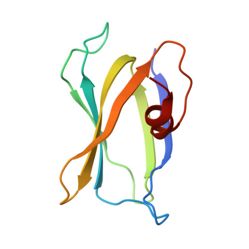
    Bacteriophages must recognize host receptors and penetrate the host cell envelope to initiate infection. How the classic phage λ initiates infection is not yet understood. Here, we combine cryo-electron microscopy and tomography to visualize infection initiation by Ur-λ, the original λ isolate that uses side fibers to adsorb rapidly to Escherichia coli . We determine the structure of Ur-λ, resolving the full-length central and side fibers, thus providing a structural basis for host recognition. We show that Ur-λ contains six copies of its tape measure protein. We capture intermediates of the tail tip complex during infection initiation, revealing how extensive conformational changes enable adsorption, and visualize the trans-envelope channel required for genome ejection.


  • Organizational Affiliation
    • Department of Microbial Pathogenesis, Yale School of Medicine, New Haven, CT, USA.

Macromolecules
Find similar proteins by:  (by identity cutoff)  |  3D Structure
Entity ID: 1
MoleculeChains Sequence LengthOrganismDetailsImage
Tail fiber protein94Escherichia phage LambdaMutation(s): 0 
Gene Names: stflambdap27
UniProt
Find proteins for P03764 (Escherichia phage lambda)
Explore P03764 
Go to UniProtKB:  P03764
Entity Groups  
Sequence Clusters30% Identity50% Identity70% Identity90% Identity95% Identity100% Identity
UniProt GroupP03764
Sequence Annotations
Expand
  • Reference Sequence
Experimental Data & Validation

Experimental Data

  • Method: ELECTRON MICROSCOPY
  • Resolution: 3.31 Å
  • Aggregation State: PARTICLE 
  • Reconstruction Method: SINGLE PARTICLE 
EM Software:
TaskSoftware PackageVersion
MODEL REFINEMENTPHENIX1.21_5190
RECONSTRUCTIONcryoSPARC

Structure Validation

View Full Validation Report


Entry History & Funding Information

Deposition Data


Funding OrganizationLocationGrant Number
National Institutes of Health/National Institute of General Medical Sciences (NIH/NIGMS)United States124378
National Institutes of Health/National Institute of General Medical Sciences (NIH/NIGMS)United States110243

Revision History  (Full details and data files)

  • Version 1.0: 2025-11-26
    Type: Initial release