9ME8 | pdb_00009me8

Co-crystal structure of maltose binding protein (MBP)-human SENP3 fusion protein in complex with PELP1 peptide

  • Classification: HYDROLASE
  • Organism(s): Homo sapiens
  • Expression System: Escherichia coli
  • Mutation(s): Yes 

  • Deposited: 2024-12-06 Released: 2025-08-06 
  • Deposition Author(s): Kaminski, A.M., Gordon, J., Pedersen, L.C., Stanley, R.E.
  • Funding Organization(s): National Institutes of Health/National Institute of Environmental Health Sciences (NIH/NIEHS), Department of Energy (DOE, United States), National Institutes of Health/National Institute of General Medical Sciences (NIH/NIGMS)

Experimental Data Snapshot

  • Method: X-RAY DIFFRACTION
  • Resolution: 2.93 Å
  • R-Value Free: 
    0.232 (Depositor), 0.230 (DCC) 
  • R-Value Work: 
    0.202 (Depositor), 0.202 (DCC) 
  • R-Value Observed: 
    0.203 (Depositor) 

Starting Model: in silico
View more details

wwPDB Validation   3D Report Full Report


This is version 1.0 of the entry. See complete history


Literature

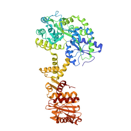
PELP1 coordinates the modular assembly and enzymatic activity of the rixosome complex.

Gordon, J.Kaminski, A.M.Bommu, S.R.Skrajna, A.Petrovich, R.M.Pedersen, L.C.McGinty, R.K.Warren, A.J.Stanley, R.E.

(2025) Sci Adv 11: eadw4603-eadw4603

  • DOI: https://doi.org/10.1126/sciadv.adw4603
  • Primary Citation of Related Structures:  
    9ME8

  • PubMed Abstract: 

    The rixosome is a large multisubunit complex that initiates RNA decay during critical nuclear transactions including ribosome assembly and heterochromatin maintenance. The overall architecture of the complex remains undefined because several subunits contain intrinsically disordered regions (IDRs). Here, we combined structural and functional approaches to establish PELP1 as the central scaffold of the rixosome upon which the enzymatic subunits modularly assemble. The C-terminal half of PELP1 is composed of a proline-rich IDR that mediates association with the AAA-ATPase MDN1, histones, and the SUMO-specific protease SENP3. The PELP1 IDR contains a glutamic acid-rich region that we establish can chaperone the histone octamer in vitro. Last, the x-ray structure of a small linear motif (SLiM) from the PELP IDR bound to SENP3 reveals how PELP1 allosterically activates SUMO protease activity. This work provides an integrated structural model for understanding the rixosome's dynamic architecture and how it modularly coordinates several cellular functions.


  • Organizational Affiliation
    • Molecular and Cellular Biology Laboratory, National Institute of Environmental Health Sciences, National Institutes of Health, Department of Health and Human Services, Research Triangle Park, NC 27709, USA.

Macromolecules
Find similar proteins by:  (by identity cutoff)  |  3D Structure
Entity ID: 1
MoleculeChains Sequence LengthOrganismDetailsImage
Maltose/maltodextrin-binding periplasmic protein,Sentrin-specific protease 3635Homo sapiensMutation(s): 1 
Gene Names: malEZ5632ECs5017SENP3SSP3SUSP3
EC: 3.4.22
UniProt & NIH Common Fund Data Resources
Find proteins for Q9H4L4 (Homo sapiens)
Explore Q9H4L4 
Go to UniProtKB:  Q9H4L4
PHAROS:  Q9H4L4
GTEx:  ENSG00000161956 
Entity Groups  
Sequence Clusters30% Identity50% Identity70% Identity90% Identity95% Identity100% Identity
UniProt GroupQ9H4L4
Sequence Annotations
Expand
  • Reference Sequence

Find similar proteins by:  Sequence   |   3D Structure  

Entity ID: 2
MoleculeChains Sequence LengthOrganismDetailsImage
Proline-, glutamic acid- and leucine-rich protein 118Homo sapiensMutation(s): 0 
UniProt & NIH Common Fund Data Resources
Find proteins for Q8IZL8 (Homo sapiens)
Explore Q8IZL8 
Go to UniProtKB:  Q8IZL8
PHAROS:  Q8IZL8
GTEx:  ENSG00000141456 
Entity Groups  
Sequence Clusters30% Identity50% Identity70% Identity90% Identity95% Identity100% Identity
UniProt GroupQ8IZL8
Sequence Annotations
Expand
  • Reference Sequence
Oligosaccharides

Help

Entity ID: 3
MoleculeChains Length2D Diagram Glycosylation3D Interactions
alpha-D-glucopyranose-(1-4)-alpha-D-glucopyranose-(1-4)-alpha-D-glucopyranose-(1-4)-alpha-D-glucopyranose
C
4N/A
Glycosylation Resources
GlyTouCan:  G87171PZ
GlyCosmos:  G87171PZ
GlyGen:  G87171PZ
Small Molecules
Ligands 1 Unique
IDChains Name / Formula / InChI Key2D Diagram3D Interactions
NA
Query on NA

Download Ideal Coordinates CCD File 
D [auth A]SODIUM ION
Na
FKNQFGJONOIPTF-UHFFFAOYSA-N
Biologically Interesting Molecules (External Reference) 1 Unique
Experimental Data & Validation

Experimental Data

  • Method: X-RAY DIFFRACTION
  • Resolution: 2.93 Å
  • R-Value Free:  0.232 (Depositor), 0.230 (DCC) 
  • R-Value Work:  0.202 (Depositor), 0.202 (DCC) 
  • R-Value Observed: 0.203 (Depositor) 
Space Group: P 32 2 1
Unit Cell:
Length ( Å )Angle ( ˚ )
a = 140.97α = 90
b = 140.97β = 90
c = 119.633γ = 120
Software Package:
Software NamePurpose
PHENIXrefinement
autoPROCdata reduction
autoPROCdata scaling
PHASERphasing

Structure Validation

View Full Validation Report


Entry History & Funding Information

Deposition Data


Funding OrganizationLocationGrant Number
National Institutes of Health/National Institute of Environmental Health Sciences (NIH/NIEHS)United StatesZIA ES103247
National Institutes of Health/National Institute of Environmental Health Sciences (NIH/NIEHS)United StatesZIC ES102645
Department of Energy (DOE, United States)United StatesDE-SC0012704
National Institutes of Health/National Institute of General Medical Sciences (NIH/NIGMS)United StatesP30M133893
National Institutes of Health/National Institute of General Medical Sciences (NIH/NIGMS)United StatesKP1605010

Revision History  (Full details and data files)

  • Version 1.0: 2025-08-06
    Type: Initial release