


A novel PE hydrolase and its structural basis
Wang, Y.S., Sun, D.Y., Jia, H.H., Sun, Y.Z., Qiu, L.N.To be published.
Experimental Data Snapshot
Entity ID: 1 | |||||
|---|---|---|---|---|---|
| Molecule | Chains | Sequence Length | Organism | Details | Image |
| Alpha/beta hydrolase | 281 | Vreelandella venusta | Mutation(s): 0 Gene Names: JDS37_04195 | 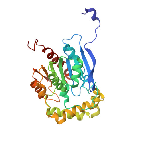 | |
UniProt | |||||
Entity Groups | |||||
| Sequence Clusters | 30% Identity50% Identity70% Identity90% Identity95% Identity100% Identity | ||||
| UniProt Group | A0AAQ0CHA8 | ||||
Sequence AnnotationsExpand | |||||
Reference Sequence | |||||
| Ligands 1 Unique | |||||
|---|---|---|---|---|---|
| ID | Chains | Name / Formula / InChI Key | 2D Diagram | 3D Interactions | |
| PEG (Subject of Investigation/LOI) Download:Ideal Coordinates CCD File | C [auth A] D [auth A] E [auth A] F [auth B] G [auth B] | DI(HYDROXYETHYL)ETHER C4 H10 O3 MTHSVFCYNBDYFN-UHFFFAOYSA-N |  | ||
| Length ( Å ) | Angle ( ˚ ) |
|---|---|
| a = 169.42 | α = 90 |
| b = 169.42 | β = 90 |
| c = 54.99 | γ = 120 |
| Software Name | Purpose |
|---|---|
| AMBER | refinement |
| AMBER | refinement |
| APEX 2 | data reduction |
| APEX 2 | data scaling |
| HKL-3000 | phasing |
| Funding Organization | Location | Grant Number |
|---|---|---|
| Other government | China | -- |


