9L5Z | pdb_00009l5z

Crystal structure of a parallel-stranded PNA duplex


Experimental Data Snapshot

  • Method: X-RAY DIFFRACTION
  • Resolution: 1.70 Å
  • R-Value Free: 
    0.290 (Depositor), 0.301 (DCC) 
  • R-Value Work: 
    0.249 (Depositor), 0.259 (DCC) 
  • R-Value Observed: 
    0.251 (Depositor) 

Starting Model: experimental
View more details

wwPDB Validation   3D Report Full Report


This is version 1.0 of the entry. See complete history


Literature

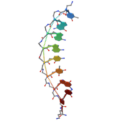
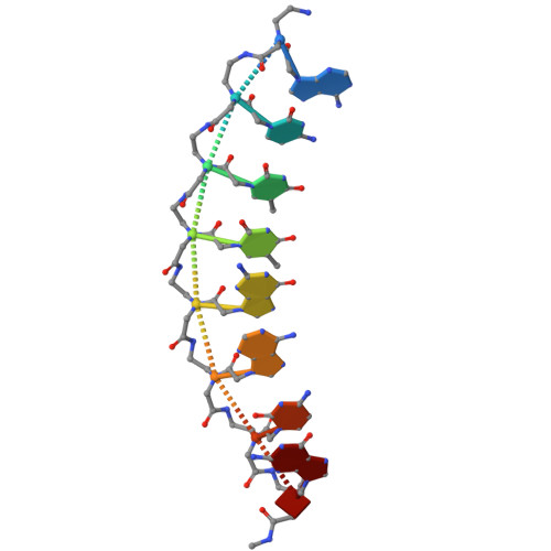
Peptide nucleic acids in parallel orientation form invasion complexes with double-stranded DNA.

Shibata, M.Sugimoto, H.Hibino, M.Shoji, O.Aiba, Y.

(2025) RSC Chem Biol 6: 1566-1575

  • DOI: https://doi.org/10.1039/d5cb00172b
  • Primary Citation of Related Structures:  
    9L5Z

  • PubMed Abstract: 

    Peptide nucleic acid (PNA) is a unique class of synthetic nucleic acids with a pseudo-peptide backbone, known for its high nucleic acid recognition capability and its ability to directly recognize double-stranded DNA (dsDNA) via the formation of a unique invasion complex. While most natural and artificial nucleic acids form duplexes in an antiparallel configuration due to the general instability of parallel configurations, PNA distinctively forms both antiparallel and parallel duplexes. In this study, we focused on this previously underexplored property of PNA to adopt a parallel duplex configuration and developed a novel double-duplex invasion strategy by leveraging the differences in thermal stability between the antiparallel and parallel orientations of PNA duplexes. Furthermore, we report the first crystal structure of a parallel PNA duplex, which was found to exhibit different structural features compared to the previously characterized antiparallel PNA duplex. This study highlights the potential of artificial nucleic acids in dsDNA recognition and demonstrates that the parallel architecture may serve as a conceptual foundation for advancing broader methodological innovations in nucleic acid research.


  • Organizational Affiliation
    • Department of Chemistry, Graduate School of Science, Nagoya University, Furo-cho Chikusa-ku Nagoya 464-8602 Japan aiba.yuichiro.f4@f.mail.nagoya-u.ac.jp +81-52-789-3557 +81-52-789-2953.

Macromolecules

Find similar proteins by:  Sequence   |   3D Structure  

Entity ID: 1
MoleculeChains Sequence LengthOrganismDetailsImage
(TPN)(GPN)(APN)(APN)(CPN)(TPN)(GPN)(CPN)(LYN)
A, C, E, G
9synthetic constructMutation(s): 0 
Sequence Annotations
Expand
  • Reference Sequence

Find similar proteins by:  Sequence   |   3D Structure  

Entity ID: 2
MoleculeChains Sequence LengthOrganismDetailsImage
(APN)(CPN)(TPN)(TPN)(GPN)(APN)(CPN)(GPN)(LYN)
B, D, F, H
9synthetic constructMutation(s): 0 
Sequence Annotations
Expand
  • Reference Sequence
Small Molecules
Modified Residues  1 Unique
IDChains TypeFormula2D DiagramParent
LYN
Query on LYN
A, C, E, G
L-PEPTIDE LINKINGC6 H16 N3 OLYS
Experimental Data & Validation

Experimental Data

  • Method: X-RAY DIFFRACTION
  • Resolution: 1.70 Å
  • R-Value Free:  0.290 (Depositor), 0.301 (DCC) 
  • R-Value Work:  0.249 (Depositor), 0.259 (DCC) 
  • R-Value Observed: 0.251 (Depositor) 
Space Group: P 1 21 1
Unit Cell:
Length ( Å )Angle ( ˚ )
a = 38.063α = 90
b = 52.867β = 94.89
c = 53.34γ = 90
Software Package:
Software NamePurpose
REFMACrefinement
Aimlessdata scaling
PHASERphasing
XDSdata reduction

Structure Validation

View Full Validation Report


Entry History & Funding Information

Deposition Data


Funding OrganizationLocationGrant Number
Japan Society for the Promotion of Science (JSPS)Japan22K05350
Japan Society for the Promotion of Science (JSPS)Japan22KJ1567

Revision History  (Full details and data files)

  • Version 1.0: 2025-12-10
    Type: Initial release