9KRQ | pdb_00009krq

Crystal structure of ZXC21 RBD


Experimental Data Snapshot

  • Method: X-RAY DIFFRACTION
  • Resolution: 3.08 Å
  • R-Value Free: 
    0.269 (Depositor), 0.273 (DCC) 
  • R-Value Work: 
    0.236 (Depositor), 0.244 (DCC) 
  • R-Value Observed: 
    0.239 (Depositor) 

Starting Model: experimental
View more details

wwPDB Validation   3D Report Full Report


Ligand Structure Quality Assessment 


This is version 1.1 of the entry. See complete history


Literature

Structural insights into the receptor-binding domain of bat coronavirus ZXC21.

Wang, C.Nan, X.Deng, Y.Fan, S.Li, X.Lan, J.

(2025) Structure 33: 1178

  • DOI: https://doi.org/10.1016/j.str.2025.04.004
  • Primary Citation of Related Structures:  
    9KRQ

  • PubMed Abstract: 

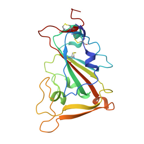
    Bat coronaviruses ZXC21 and ZC45 were discovered before the COVID-19 outbreak and share approximately 86% genome homology with SARS-CoV-2. Earlier studies indicated that ZXC21 and ZC45 may be involved in the emergence of SARS-CoV-2. However, the cell invasion mechanisms of ZXC21 and ZC45 remain unclear. Here, we determined the crystal structure of the ZXC21 receptor-binding domain (RBD) and found that the core structure shared high similarity with SARS-CoV-2, MERS-CoV, human coronavirus (HCoV)-HKU1, SARS-CoV, and HCoV-OC43 RBDs, whereas the receptor-binding motifs (RBMs) differ. We demonstrated that the ZXC21 RBD had no interaction with the human coronavirus receptors angiotensin-converting enzyme 2 (ACE2), dipeptidylpeptidase 4 (DPP4), aminopeptidase N (APN), or transmembrane serine protease 2 (TMPRSS2) by surface plasmon resonance (SPR). Moreover, the P5S-3B11 Fab can bind to the ZXC21 RBD, indicating that this SARS-CoV-2 core-targeting antibody may retain neutralizing activity toward the ZXC21 coronavirus. Our results revealed the bat coronavirus ZXC21 RBD structure, which may provide further insights into the evolution of SARS-CoV-2 and the other human beta-coronaviruses.


  • Organizational Affiliation
    • School of Biomedical Sciences, Hunan University, Changsha, Hunan, China.

Macromolecules
Find similar proteins by:  (by identity cutoff)  |  3D Structure
Entity ID: 1
MoleculeChains Sequence LengthOrganismDetailsImage
Spike glycoproteinA [auth B],
B [auth A]
181Bat coronavirusMutation(s): 0 
UniProt
Find proteins for A0A2R3SUW9 (Bat SARS-like coronavirus)
Explore A0A2R3SUW9 
Go to UniProtKB:  A0A2R3SUW9
Entity Groups  
Sequence Clusters30% Identity50% Identity70% Identity90% Identity95% Identity100% Identity
UniProt GroupA0A2R3SUW9
Glycosylation
Glycosylation Sites: 1
Sequence Annotations
Expand
  • Reference Sequence
Small Molecules
Ligands 1 Unique
IDChains Name / Formula / InChI Key2D Diagram3D Interactions
NAG (Subject of Investigation/LOI)
Query on NAG

Download Ideal Coordinates CCD File 
C [auth B]2-acetamido-2-deoxy-beta-D-glucopyranose
C8 H15 N O6
OVRNDRQMDRJTHS-FMDGEEDCSA-N
Experimental Data & Validation

Experimental Data

  • Method: X-RAY DIFFRACTION
  • Resolution: 3.08 Å
  • R-Value Free:  0.269 (Depositor), 0.273 (DCC) 
  • R-Value Work:  0.236 (Depositor), 0.244 (DCC) 
  • R-Value Observed: 0.239 (Depositor) 
Space Group: P 21 21 21
Unit Cell:
Length ( Å )Angle ( ˚ )
a = 44.163α = 90
b = 73.772β = 90
c = 117.835γ = 90
Software Package:
Software NamePurpose
PHENIXrefinement
HKL-2000data reduction
HKL-2000data scaling
PHENIXphasing

Structure Validation

View Full Validation Report


Ligand Structure Quality Assessment 


Entry History & Funding Information

Deposition Data


Funding OrganizationLocationGrant Number
Not funded--

Revision History  (Full details and data files)

  • Version 1.0: 2025-05-14
    Type: Initial release
  • Version 1.1: 2025-07-23
    Changes: Database references