Fluorogen-Activating Human Serum Albumin for Mitochondrial Nanoscale Imaging.
Fang, B., Bai, H., Zhang, J., Shi, M., Ge, Y., Wang, L., Li, P., Ding, Y., Zhang, S., Zhang, C., Qu, Y., Zhang, D., Peng, B., Chen, X., Li, L., Huang, W.(2025) Adv Mater 37: e2501849-e2501849
- PubMed: 40420673 Search on PubMed
- DOI: https://doi.org/10.1002/adma.202501849
- Primary Citation Related Structures:
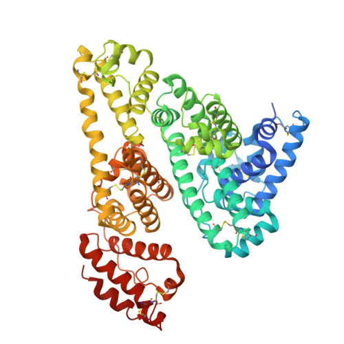
9KQK - PubMed Abstract:
Fluorescence nanoscopy of living cells employs contrast agents to reveal intrinsic correlations between mitochondrial dynamics and functions at the molecular level. However, regular mitochondrial fluorophores usually present poor photostability, low brightness, non-specific inhibitory effects, high phototoxicity, and rapid photobleaching, which have hindered the use of these tools to capture the intricate dynamic features of mitochondria. Herein, we engineered a fluorogen-activating protein (FAP), AmpHecy@HSA, a non-covalent self-assembly of HSA and amphiphilic hemicyanine (AmpHecy) fluorophore, with exceptional cell permeability, long-lasting photostability, high brightness/fluorogenicity, and minimal phototoxicity. Crystallography and femtosecond transient absorption spectroscopy techniques were combined to elucidate the structural and mechanistic intricacies of fluorescence activation. These findings revealed that fluorophore photoactivation happens through the molecular conformation-induced intramolecular charge transfer, whose kinetics is mainly determined by the hydrophobic interaction between the fluorophore and nearby amino acids. This aligns with classical molecular dynamics simulations and excited-state conformation quantum mechanics. It was further demonstrated that AmpHecy@HSA can be used for super-resolved images of mitochondria within living cells without apparent phototoxicity. This work expands the fluorescent toolkit based on FAP engineering for studying live-cell mitochondrial morphology and function, advancing the fields of chemistry and biomedicine.
- State Key Laboratory of Flexible Electronics (LoFE) & Institute of Flexible Electronics (IFE), Xiamen University, Xiamen, 361102, China.
Organizational Affiliation: