9KO3 | pdb_00009ko3

Crystal structure of Agrobacterium tumefaciens PmtA with SAH


Experimental Data Snapshot

  • Method: X-RAY DIFFRACTION
  • Resolution: 1.95 Å
  • R-Value Free: 
    0.227 (Depositor), 0.227 (DCC) 
  • R-Value Work: 
    0.187 (Depositor), 0.189 (DCC) 
  • R-Value Observed: 
    0.189 (Depositor) 

Starting Model: in silico
View more details

wwPDB Validation 3D Report Full Report

Validation slider image for 9KO3

Ligand Structure Quality Assessment 


This is version 1.1 of the entry. See complete history

Literature

Structural basis for phosphatidylcholine synthesis by bacterial phospholipid N-methyltransferases.

Watanabe, Y.Kumeta, H.Watanabe, S.

(2025) J Biological Chem 301: 108507-108507

  • DOI: https://doi.org/10.1016/j.jbc.2025.108507
  • Primary Citation Related Structures: 
    9KO3, 9KO5

  • PubMed Abstract: 

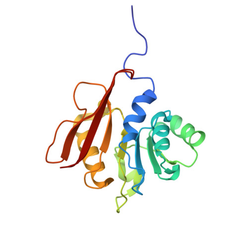
    In phosphatidylcholine (PC)-containing bacteria, PC is synthesized by phospholipid N-methyltransferases (Pmts) and plays an important role in the interactions between symbiotic and pathogenic bacteria and their eukaryotic host cells. Pmts catalyze the SAM-dependent three methylation reactions of the head group of phosphatidylethanolamine (PE) to form PC through monomethyl PE and dimethyl PE. However, the precise molecular mechanisms underlying PC biosynthesis by PmtA remain largely unclear, owing to the lack of structural information. Here, we determined the crystal structures of Agrobacterium tumefaciens Pmt (AtPmtA) in complex with SAH or 5'-methylthioadenosine. Crystal structures and NMR analysis revealed the binding mode of AtPmtA to SAH in solution. Structure-based mutational analyses showed that a conserved tyrosine residue in the substrate-binding groove is involved in methylation. Furthermore, we showed that differences in substrate specificity among Pmt homologs were determined by whether the amino acid residues comprising the substrate-binding groove were isoleucine or phenylalanine. These findings provide a structural basis for understanding the mechanisms underlying Pmts-mediated PC biosynthesis.


  • Organizational Affiliation
    • Faculty of Science, Yamagata University, Yamagata, Japan. Electronic address: yasunori@sci.kj.yamagata-u.ac.jp.

Macromolecule Content 

  • Total Structure Weight: 77.81 kDa 
  • Atom Count: 5,891 
  • Modeled Residue Count: 666 
  • Deposited Residue Count: 696 
  • Unique protein chains: 1

Macromolecules

Find similar proteins by:|  3D Structure
Entity ID: 1
MoleculeChains  Sequence LengthOrganismDetailsImage
Methyltransferase
A, B, C, D
174Agrobacterium fabrum str. C58Mutation(s): 0 
Gene Names: Atu0300
UniProt
Find proteins for A9CKE9 (Agrobacterium fabrum (strain C58 / ATCC 33970))
Explore A9CKE9 
Go to UniProtKB:  A9CKE9
Entity Groups
Sequence Clusters30% Identity50% Identity70% Identity90% Identity95% Identity100% Identity
UniProt GroupA9CKE9
Sequence Annotations
Expand
Reference Sequence

Experimental Data & Validation

Experimental Data

  • Method: X-RAY DIFFRACTION
  • Resolution: 1.95 Å
  • R-Value Free:  0.227 (Depositor), 0.227 (DCC) 
  • R-Value Work:  0.187 (Depositor), 0.189 (DCC) 
  • R-Value Observed: 0.189 (Depositor) 
Space Group: P 1 21 1
Unit Cell:
Length ( Å )Angle ( ˚ )
a = 69.942α = 90
b = 76.378β = 105.83
c = 87.185γ = 90
Software Package:
Software NamePurpose
PHENIXrefinement
XDSdata scaling
XDSdata reduction
PHASERphasing

Structure Validation

View Full Validation Report


Ligand Structure Quality Assessment 


Entry History 

& Funding Information

Deposition Data

  • Released Date: 2025-04-23 
  • Deposition Author(s): Watanabe, Y.

Funding OrganizationLocationGrant Number
Japan Society for the Promotion of Science (JSPS)Japan22K06096
Japan Society for the Promotion of Science (JSPS)Japan20K15734

Revision History  (Full details and data files)

  • Version 1.0: 2025-04-23
    Type: Initial release
  • Version 1.1: 2026-05-06
    Changes: Database references