9KL2 | pdb_00009kl2

Crystal structure of Pleurocybella porrigens lectin (PPL) in complex with GalNAc


Experimental Data Snapshot

  • Method: X-RAY DIFFRACTION
  • Resolution: 2.00 Å
  • R-Value Free: 
    0.249 (Depositor), 0.249 (DCC) 
  • R-Value Work: 
    0.228 (Depositor), 0.228 (DCC) 
  • R-Value Observed: 
    0.230 (Depositor) 

Starting Model: in silico
View more details

wwPDB Validation   3D Report Full Report


Ligand Structure Quality Assessment 


This is version 1.2 of the entry. See complete history


Literature

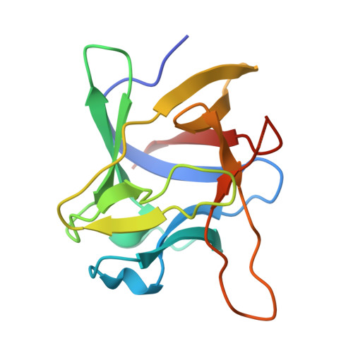
Crystal and Cryo-EM structure of PPL, a novel hexameric R-type lectin from the poisonous mushroom Pleurocybella porrigens.

Adachi, D.Ishimoto, N.Mizutani, K.Takahashi, K.Kubota, R.Kawabata, H.Park, S.Y.Vandebroek, L.Voet, A.R.D.Yamada, M.Ozeki, Y.Fujii, Y.Fujita, H.Tame, J.R.H.Kamata, K.

(2025) Glycobiology 36

  • DOI: https://doi.org/10.1093/glycob/cwaf082
  • Primary Citation of Related Structures:  
    9KL2, 9KL3

  • PubMed Abstract: 

    Pleurocybella porrigens is a mushroom that grows widely around the temperate northern hemisphere, and was once considered edible, especially in Japan. Following a number of deaths in 2004, investigations revealed the presence of various toxins, including a lectin (PPL) that apparently survives cooking and enters the bloodstream via the stomach. We have cloned PPL and solved its structure by X-ray crystallography and cryo-EM. We report the sugar binding properties of this β-trefoil lectin, which has a novel hexameric structure.


  • Organizational Affiliation
    • Graduate School of Medical Life Science, Yokohama City University, Japan.

Macromolecules
Find similar proteins by:  (by identity cutoff)  |  3D Structure
Entity ID: 1
MoleculeChains Sequence LengthOrganismDetailsImage
Lectin
A, B, C
140Pleurocybella porrigensMutation(s): 0 
UniProt
Find proteins for B6ZK51 (Pleurocybella porrigens)
Explore B6ZK51 
Go to UniProtKB:  B6ZK51
Entity Groups  
Sequence Clusters30% Identity50% Identity70% Identity90% Identity95% Identity100% Identity
UniProt GroupB6ZK51
Sequence Annotations
Expand
  • Reference Sequence
Experimental Data & Validation

Experimental Data

  • Method: X-RAY DIFFRACTION
  • Resolution: 2.00 Å
  • R-Value Free:  0.249 (Depositor), 0.249 (DCC) 
  • R-Value Work:  0.228 (Depositor), 0.228 (DCC) 
  • R-Value Observed: 0.230 (Depositor) 
Space Group: C 2 2 21
Unit Cell:
Length ( Å )Angle ( ˚ )
a = 52.762α = 90
b = 221.166β = 90
c = 112.209γ = 90
Software Package:
Software NamePurpose
PHENIXrefinement
Aimlessdata scaling
XDSdata reduction
PHASERphasing

Structure Validation

View Full Validation Report


Ligand Structure Quality Assessment 


Entry History & Funding Information

Deposition Data


Funding OrganizationLocationGrant Number
Institute for Fermentation, OsakaJapanY-2023-2-046

Revision History  (Full details and data files)

  • Version 1.0: 2025-11-19
    Type: Initial release
  • Version 1.1: 2025-12-17
    Changes: Database references
  • Version 1.2: 2025-12-31
    Changes: Database references