Structural basis of plant organelle C-to-U RNA editing by PPR-DYW proteins.
Teramoto, T., Urushihara, R., Aoyama, R., Okada, A., Ichinose, M., Yagi, Y., Nakamura, T., Gutmann, B., Kakuta, Y.(2026) Nat Commun
- PubMed: 42045200 Search on PubMed
- DOI: https://doi.org/10.1038/s41467-026-72391-y
- Primary Citation Related Structures:
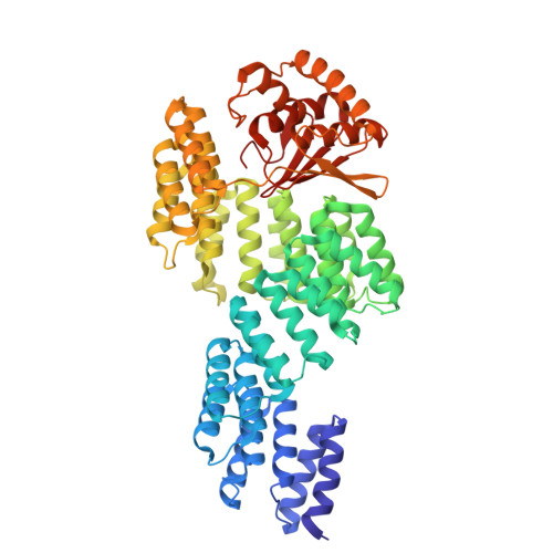
9KFX, 9KFY - PubMed Abstract:
Plants possess a unique C-to-U RNA editing mechanism mediated by PPR-DYW proteins, wherein the PPR domain recognizes specific RNA sequences while the DYW deaminase domain precisely edits the target C base-a process essential for functional protein expression in plant chloroplasts and mitochondria. The coordination of these two domains is considered crucial for precise RNA editing. In nature, this site-specific and precise base editing by PPR-DYW proteins distinguishes them from other base-editing deaminases. However, the absence of structures containing both PPR and DYW domains has limited our understanding of the precise RNA-editing mechanism of PPR-DYW proteins. Here, we present crystal structures of the consensus PPR-DYW (consPPR-DYW) protein, a representative of the PPR-DYW proteins, in both RNA-free and target RNA-bound states. Comparison between these states demonstrates domain movements upon target RNA binding, whereby the PPR domain accommodates the upstream sequence of the target C base in the proper conformation for editing while the DYW domain is optimally positioned for precise C-to-U conversion. These results, combined with comprehensive biochemical analyses, provide the foundation for a mechanistic model that explains the coordinated action of the PPR and DYW domains in achieving precise C-to-U editing.
- Laboratory of Biophysical Chemistry, Department of Bioscience and Biotechnology, Faculty of Agriculture, Kyushu University, Fukuoka, Japan. teramotot@agr.kyushu-u.ac.jp.
Organizational Affiliation: