9KEF | pdb_00009kef

Crystal strucutre of RABV-G in complex with SOJB-Fab complex


Experimental Data Snapshot

  • Method: X-RAY DIFFRACTION
  • Resolution: 3.40 Å
  • R-Value Free: 
    0.264 (Depositor), 0.275 (DCC) 
  • R-Value Work: 
    0.217 (Depositor), 0.240 (DCC) 
  • R-Value Observed: 
    0.220 (Depositor) 

Starting Model: experimental
View more details

wwPDB Validation   3D Report Full Report


Ligand Structure Quality Assessment 


This is version 1.0 of the entry. See complete history


Literature

Molecular basis of two broad-spectrum antibodies neutralizing rabies virus and other phylogroup-I lyssaviruses by blocking structural transition between the pleckstrin-homology and fusion domains in the glycoprotein.

Yang, F.Zhai, L.Yin, K.Lin, S.Yang, J.Ye, F.Chen, Z.Shu, S.Yu, Y.Guo, L.He, B.Wang, W.Ye, H.Cao, Y.Gao, J.Lu, G.

(2025) Int J Biol Macromol 308: 142570-142570

  • DOI: https://doi.org/10.1016/j.ijbiomac.2025.142570
  • Primary Citation of Related Structures:  
    9KEF, 9KEP, 9KFB

  • PubMed Abstract: 

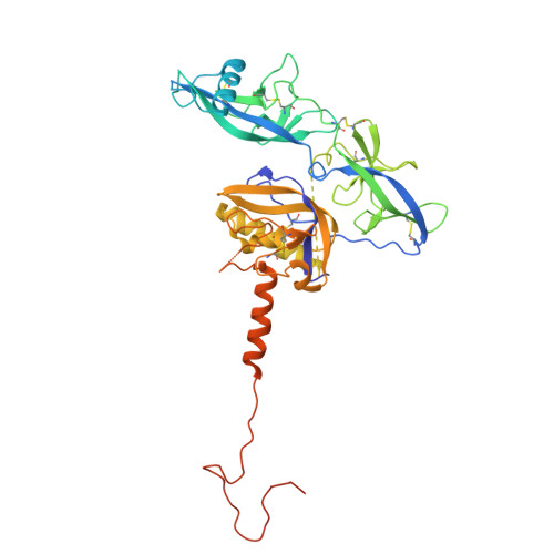
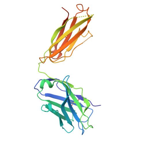
    Rabies virus (RABV), the prototype species of the Lyssavirus genus, causes the lethal disease of rabies. Rabies can be efficiently prevented with post-exposure prophylaxis. The RABV glycoprotein (RABV-G) is an essential factor mediating virus entry and the major target of neutralizing antibodies. Here, we report the crystal structures of two neutralizing monoclonal antibodies (NM57 and SOJB) in complex with RABV-G. The two antibodies recognize highly overlapped epitopes involving both the pleckstrin-homology domain (PHD) and the junction between PHD and the fusion domain (FD). Our pseudovirus neutralization assay further shows that both antibodies could neutralize a majority of the lyssaviruses in phylogroup-I. Via sequence comparison and structural characterization, we identify two residues located at positions 226 and 231 in PHD, as key determinants for antibody recognition, which is further corroborated by mutagenesis analyses. Finally, detailed structural analyses reveal that NM57 and SOJB would lock the PHD/FD local structure in the pre-fusion-like state, thereby inhibiting viral infection by blocking structural transitions of RABV-G essential for membrane fusion. Taken together, these results provide a mechanistic glimpse into the molecular basis for broad neutralization of phylogroup-I lyssaviruses by NM57 and SOJB, which should be able to facilitate the development of monoclonal antibodies and vaccines.


  • Organizational Affiliation
    • Department of Emergency Medicine, State Key Laboratory of Biotherapy, West China Hospital, Sichuan University, Chengdu, Sichuan 610041, China. Electronic address: yfanli16@qq.com.

Macromolecules
Find similar proteins by:  (by identity cutoff)  |  3D Structure
Entity ID: 1
MoleculeChains Sequence LengthOrganismDetailsImage
RABV-G439Rabies virus CVS-11Mutation(s): 0 
Entity Groups  
Sequence Clusters30% Identity50% Identity70% Identity90% Identity95% Identity100% Identity
Glycosylation
Glycosylation Sites: 1
Sequence Annotations
Expand
  • Reference Sequence
Find similar proteins by:  (by identity cutoff)  |  3D Structure
Entity ID: 2
MoleculeChains Sequence LengthOrganismDetailsImage
SOJB-Fab HchainB [auth H]239Homo sapiensMutation(s): 0 
Entity Groups  
Sequence Clusters30% Identity50% Identity70% Identity90% Identity95% Identity100% Identity
Sequence Annotations
Expand
  • Reference Sequence
Find similar proteins by:  (by identity cutoff)  |  3D Structure
Entity ID: 3
MoleculeChains Sequence LengthOrganismDetailsImage
SOJB-Fab LchianC [auth L]215Homo sapiensMutation(s): 0 
Entity Groups  
Sequence Clusters30% Identity50% Identity70% Identity90% Identity95% Identity100% Identity
Sequence Annotations
Expand
  • Reference Sequence
Small Molecules
Ligands 1 Unique
IDChains Name / Formula / InChI Key2D Diagram3D Interactions
NAG
Query on NAG

Download Ideal Coordinates CCD File 
D [auth A]2-acetamido-2-deoxy-beta-D-glucopyranose
C8 H15 N O6
OVRNDRQMDRJTHS-FMDGEEDCSA-N
Experimental Data & Validation

Experimental Data

  • Method: X-RAY DIFFRACTION
  • Resolution: 3.40 Å
  • R-Value Free:  0.264 (Depositor), 0.275 (DCC) 
  • R-Value Work:  0.217 (Depositor), 0.240 (DCC) 
  • R-Value Observed: 0.220 (Depositor) 
Space Group: C 2 2 21
Unit Cell:
Length ( Å )Angle ( ˚ )
a = 84.184α = 90
b = 154.417β = 90
c = 191.248γ = 90
Software Package:
Software NamePurpose
PHENIXrefinement
HKL-3000data scaling
PHASERphasing
DENZOdata reduction

Structure Validation

View Full Validation Report


Ligand Structure Quality Assessment 


Entry History & Funding Information

Deposition Data


Funding OrganizationLocationGrant Number
Not funded--

Revision History  (Full details and data files)

  • Version 1.0: 2025-04-23
    Type: Initial release