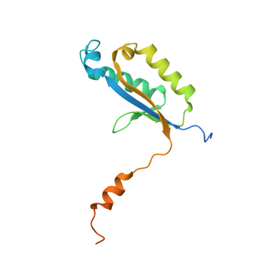
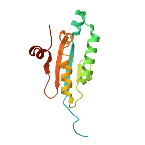
Novel type II toxin-antitoxin systems with VapD-like proteins.
Gilep, K., Bikmetov, D., Popov, A., Rusanova, A., Tagami, S., Dubiley, S., Severinov, K.(2025) mBio 16: e0000325-e0000325
- PubMed: 40052803 Search on PubMed
- DOI: https://doi.org/10.1128/mbio.00003-25
- Primary Citation Related Structures:
9KAN - PubMed Abstract:
Type II toxin-antitoxin (TA) systems are widespread in prokaryotes. They consist of neighboring genes encoding two small proteins: a toxin that inhibits a critical cellular process and an antitoxin that binds to and neutralizes the toxin. The VapD nuclease and the VapX antitoxin comprise a type II TA system that contributes to the virulence of the human pathogen Haemophilus influenzae . We analyzed the diversity and evolution of VapD-like proteins. By examining loci adjacent to genes coding for VapD-like proteins, we identified two novel families of antitoxins, which we named VapY and VapW. VapD toxins cognate to novel antitoxins induce the SOS response when overproduced, suggesting they target cellular processes related to genomic DNA integrity, maintenance, or replication. Though VapY has no sequence similarity to VapX, they share the same SH3 fold characterized by the five anti-parallel β sheets that form a barrel. VapW is a homolog of VapD without conserved catalytic residues required for nuclease activity. The crystal structure of the VapD-VapW complex reveals that VapW lacks the dimerization interface essential for the catalytic activity of VapD but retains the second interaction interface that enables VapD hexamerization. This allows VapW to bind VapD in the same manner that VapD dimers bind to each other in hexamers. Thus, though the VapD catalytic cleft remains accessible in the VapD-VapW complex, VapW may disrupt VapD oligomerization. To our knowledge, VapWD provides a unique example of TA systems evolution when a toxin loses its activity and becomes an antitoxin to itself. Genes encoding virulence-associated protein D (VapD) homologs are found in many pathogens such as Helicobacter pylori , Haemophilus influenzae , and Xylella fastidiosa . There are many indications that VapD proteins contribute to virulence, even though the exact mechanism is not known. VapD proteins are either encoded by stand-alone genes or form toxin-antitoxin pairs with VapX. We performed a comprehensive census of vapD-like genes and found two new antitoxins, VapW and VapY. The VapW antitoxins are catalytically inactivated variants of VapD, revealing a new evolutionary mechanism for the appearance of toxin-antitoxin pairs.
- Center for Precision Genome Editing and Genetic Technologies for Biomedicine Institute of Gene Biology, Russian Academy of Sciences, Moscow, Russia.
Organizational Affiliation: