9K2J | pdb_00009k2j

X-ray crystal structure of 3-hydroxyisobutyrate dehydrogenase


Experimental Data Snapshot

  • Method: X-RAY DIFFRACTION
  • Resolution: 2.88 Å
  • R-Value Free: 
    0.240 (Depositor), 0.240 (DCC) 
  • R-Value Work: 
    0.204 (Depositor), 0.207 (DCC) 
  • R-Value Observed: 
    0.206 (Depositor) 

Starting Model: experimental
View more details

wwPDB Validation   3D Report Full Report


Ligand Structure Quality Assessment 


This is version 1.0 of the entry. See complete history


Literature

Mirror substrates specificity of a 2, 3-dihydroxypropanesulfonate degrading enzyme in sulfate-reducing bacteria.

Ma, X.Wang, H.Liu, L.Dang, H.Tang, K.

(2025) Int J Biol Macromol 306: 141806-141806

  • DOI: https://doi.org/10.1016/j.ijbiomac.2025.141806
  • Primary Citation of Related Structures:  
    9K2J

  • PubMed Abstract: 

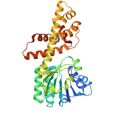
    Ubiquitous R- and S-enantiomers of 2,3-dihydroxypropanesulfonate (DHPS), organic sulfur compounds produced by photosynthetic organisms, serve as common nutrient and energy sources for specific bacteria. While most known DHPS-degrading enzymes exhibit enantioselectivity, this study introduces a unique dehydrogenase, DhpA from the sulfate-reducing bacterium Desulfovibrio sp. DF1, capable of efficiently metabolizing both R- and S-DHPS to 3-sulfolactaldehyde (SLA). The crystal structure of DhpA reveals a conserved binding pocket that recognizes the sulfonate group of DHPS through interactions with Lys123, Ser174, and Asn175. The catalytic mechanism of the enzyme involves the oxidation of the C3-OH group of both enantiomers, facilitated by the Lys171. The mutation of Lys171 significantly diminishes activity, confirming its critical role in catalysis. Based on biochemical and genetic analyses, this study proposes a chiral DHPS degradation pathway in bacteria. This study reveals the unique enantiomeric selectivity of DhpA, expanding our understanding of the bacterial metabolism of chiral molecules.


  • Organizational Affiliation
    • State Key Laboratory of Marine Environmental Science, Fujian Key Laboratory of Marine Carbon Sequestration, College of Ocean and Earth Science, Xiamen University, Xiamen, China.

Macromolecules
Find similar proteins by:  (by identity cutoff)  |  3D Structure
Entity ID: 1
MoleculeChains Sequence LengthOrganismDetailsImage
3-hydroxyisobutyrate dehydrogenase
A, B, C
286Desulfovibrio sp.Mutation(s): 0 
Entity Groups  
Sequence Clusters30% Identity50% Identity70% Identity90% Identity95% Identity100% Identity
Sequence Annotations
Expand
  • Reference Sequence
Experimental Data & Validation

Experimental Data

  • Method: X-RAY DIFFRACTION
  • Resolution: 2.88 Å
  • R-Value Free:  0.240 (Depositor), 0.240 (DCC) 
  • R-Value Work:  0.204 (Depositor), 0.207 (DCC) 
  • R-Value Observed: 0.206 (Depositor) 
Space Group: I 41 2 2
Unit Cell:
Length ( Å )Angle ( ˚ )
a = 289.16α = 90
b = 289.16β = 90
c = 65.87γ = 90
Software Package:
Software NamePurpose
PHENIXrefinement
PHENIXmodel building
Aimlessdata scaling
PHASERphasing

Structure Validation

View Full Validation Report


Ligand Structure Quality Assessment 


Entry History & Funding Information

Deposition Data

  • Released Date: 2025-03-19 
  • Deposition Author(s): Tang, K.

Funding OrganizationLocationGrant Number
National Natural Science Foundation of China (NSFC)China42188102, 42141003, 42076160

Revision History  (Full details and data files)

  • Version 1.0: 2025-03-19
    Type: Initial release