SAMD12 as a Master Regulator of MAP4Ks by Decoupling Kinases From the CNKSR2 Scaffold.
Pan, W., Lin, Z., Chen, S., Li, J., Wang, Y., Chen, K., Zhang, M.(2025) J Mol Biology 437: 169034-169034
- PubMed: 40010432 Search on PubMed
- DOI: https://doi.org/10.1016/j.jmb.2025.169034
- Primary Citation Related Structures:
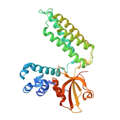
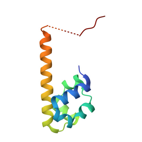
9K1L - PubMed Abstract:
The MAP4K member TNIK and the multi-domain scaffold protein CNKSR2, both of which are clustered at neuronal synapses, interact with each other and are closely associated with neurodevelopmental disorders, although the mechanism underlying their interaction is unclear. In this study, we characterized the interaction mechanisms between MAP4K kinases (MAP4K4, MINK1 and TNIK) and the CNKSR1/2/3 scaffold proteins, and discovered that SAMD12, a familial adult myoclonic epilepsy disease gene product, or its close homolog SAMD10, binds to CNKSR1/2/3 with exceptionally strong affinities and can quantitatively displace MAP4K from CNKSR1/2/3 scaffolds. Additionally, we demonstrated that CNKSR2 acts as both a scaffold and an activator of TNIK during neuronal synapse development. Ectopic expression of SAMD12 can effectively alter synapse development, likely by inhibiting TNIK activity through the dissociation of the kinase from CNKSR2. Our findings may have broad implications on the roles of MAP4Ks and CNKSR1/2/3 in the nervous system and in other tissues under physiological and pathophysiological processes.
- Shenzhen Key Laboratory of Biomolecular Assembling and Regulation, School of Life Sciences, Southern University of Science and Technology, Shenzhen 518055, China.
Organizational Affiliation: