9JSP | pdb_00009jsp

inactive NbaSPARDA complexes


Experimental Data Snapshot

  • Method: ELECTRON MICROSCOPY
  • Resolution: 3.34 Å
  • Aggregation State: PARTICLE 
  • Reconstruction Method: SINGLE PARTICLE 

wwPDB Validation 3D Report Full Report

Validation slider image for 9JSP

This is version 1.2 of the entry. See complete history

Literature

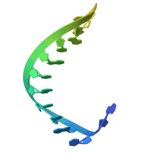
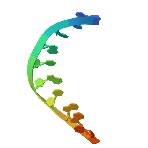
Target DNA-induced filament formation and nuclease activation of SPARDA complex.

Wang, F.Xu, H.Zhang, C.Xue, J.Li, Z.

(2025) Cell Res 35: 510-519

  • DOI: https://doi.org/10.1038/s41422-025-01100-z
  • Primary Citation Related Structures: 
    9JSB, 9JSP, 9JSZ, 9JT2

  • PubMed Abstract: 

    The short Argonaute-based bacterial defense system, SPARDA (Short Prokaryotic Argonaute and DNase/RNase-APAZ), utilizes guide RNA to target invading complementary DNA and exhibits collateral nuclease activity, leading to cell death or dormancy. However, its detailed mechanisms remain poorly understood. In this study, we investigated the SPARDA system from Novosphingopyxis baekryungensis (NbaSPARDA) and discovered an unexpected filament configuration upon target DNA binding, which strongly correlated with collateral nuclease activity. Filament formation and nuclease activation require a guide-target heteroduplex of sufficient length with perfect complementarity at the central region. A series of cryo-EM structures of NbaSPARDA complexes, loaded with guide RNA, target DNA of varying lengths, and substrate ssDNA, were determined at ~3.0 Å resolution. Structural analyses indicated that guide RNA binding induces dimerization of the NbaSPARDA complex, while target DNA engagement disrupts this dimerization. Further propagation of the guide-target heteroduplex triggers filament formation through a checkpoint mechanism. The NbaSPARDA filament consists of a backbone formed by interlocking short Argonaute proteins, with an inner layer composed of DREN nuclease domains. Filament formation leads to tetramerization of the monomeric DREN nuclease domain, activating its collateral nuclease activity against environmental nucleic acids - a feature leveraged for molecular diagnostics. For bacteria heterologously expressing the NbaSPARDA system, defense against invading bacteriophages and plasmids relies on filament formation. Collectively, these findings illustrate the detailed working mechanism of the NbaSPARDA complex and highlight the importance of its filament formation in host defense.


  • Organizational Affiliation
    • State Key Laboratory of Biocatalysis and Enzyme Engineering, School of Life Sciences, Hubei University, Wuhan, Hubei, China.

Macromolecule Content 

  • Total Structure Weight: 115.33 kDa 
  • Atom Count: 5,862 
  • Modeled Residue Count: 695 
  • Deposited Residue Count: 960 
  • Unique protein chains: 2
  • Unique nucleic acid chains: 2

Macromolecules


Find similar proteins by:|  3D Structure
Entity ID: 1
MoleculeChains  Sequence LengthOrganismDetailsImage
Ago485Novosphingopyxis baekryungensis DSM 16222Mutation(s): 0 
UniProt
Find proteins for A0A1L3JCL0 (Sphingorhabdus lutea)
Explore A0A1L3JCL0 
Go to UniProtKB:  A0A1L3JCL0
Entity Groups
Sequence Clusters30% Identity50% Identity70% Identity90% Identity95% Identity100% Identity
UniProt GroupA0A1L3JCL0
Sequence Annotations
Expand
Reference Sequence
Find similar proteins by:|  3D Structure
Entity ID: 2
MoleculeChains  Sequence LengthOrganismDetailsImage
DREN-APAZ442Novosphingopyxis baekryungensis DSM 16222Mutation(s): 0 
UniProt
Find proteins for A0A1L3JCN1 (Sphingorhabdus lutea)
Explore A0A1L3JCN1 
Go to UniProtKB:  A0A1L3JCN1
Entity Groups
Sequence Clusters30% Identity50% Identity70% Identity90% Identity95% Identity100% Identity
UniProt GroupA0A1L3JCN1
Sequence Annotations
Expand
Reference Sequence
Find similar nucleic acids by:  Sequence
Entity ID: 3
MoleculeChains LengthOrganismImage
RNA (5'-R(P*AP*UP*AP*CP*UP*GP*CP*AP*CP*AP*GP*CP*UP*GP*AP*CP*GP*AP*UP*A)-3')C [auth G]20Novosphingopyxis baekryungensis DSM 16222
Sequence Annotations
Expand
Reference Sequence
Find similar nucleic acids by:  Sequence
Entity ID: 4
MoleculeChains LengthOrganismImage
DNA (5'-D(P*GP*CP*TP*GP*TP*GP*CP*AP*GP*TP*AP*TP*T)-3')D [auth T]13Novosphingopyxis baekryungensis DSM 16222
Sequence Annotations
Expand
Reference Sequence

Small Molecules

Ligands 1 Unique
IDChains Name / Formula / InChI Key2D Diagram3D Interactions
MG
(Subject of Investigation/LOI)

Query on MG


Download:Ideal Coordinates CCD File
E [auth A]MAGNESIUM ION
Mg
JLVVSXFLKOJNIY-UHFFFAOYSA-N

Experimental Data & Validation

Experimental Data

  • Method: ELECTRON MICROSCOPY
  • Resolution: 3.34 Å
  • Aggregation State: PARTICLE 
  • Reconstruction Method: SINGLE PARTICLE 
EM Software:
TaskSoftware PackageVersion
MODEL REFINEMENTPHENIX

Structure Validation

View Full Validation Report


Entry History 

& Funding Information

Deposition Data

  • Released Date: 2025-04-02 
  • Deposition Author(s): Zhuang, L.

Funding OrganizationLocationGrant Number
Not funded--

Revision History  (Full details and data files)

  • Version 1.0: 2025-04-02
    Type: Initial release
  • Version 1.1: 2025-07-16
    Changes: Data collection, Database references
  • Version 1.2: 2025-07-23
    Changes: Data collection