9JQ5 | pdb_00009jq5

Structure of human IVD in complex with FAD and isovaleryl-CoA


Experimental Data Snapshot

  • Method: ELECTRON MICROSCOPY
  • Resolution: 3.35 Å
  • Aggregation State: PARTICLE 
  • Reconstruction Method: SINGLE PARTICLE 

wwPDB Validation   3D Report Full Report


This is version 1.1 of the entry. See complete history


Literature

Structural Insights into Isovaleryl-Coenzyme A Dehydrogenase: Mechanisms of Substrate Specificity and Implications of Isovaleric Acidemia-Associated Mutations.

Ju, K.Bai, F.Xu, Y.Li, Q.Su, G.Jin, Y.Chen, H.Zhang, S.Luan, X.

(2025) Research (Wash D C) 8: 0661-0661

  • DOI: https://doi.org/10.34133/research.0661
  • Primary Citation of Related Structures:  
    9JQ3, 9JQ4, 9JQ5

  • PubMed Abstract: 

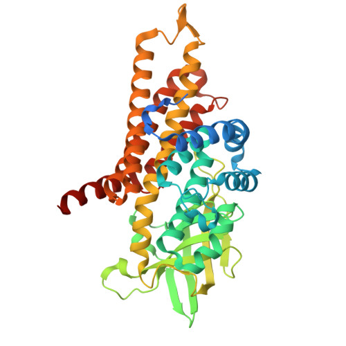
    Isovaleryl-CoA (coenzyme A) dehydrogenase (IVD) plays a pivotal role in the catabolism of leucine, converting isovaleryl-CoA to 3-methylcrotonyl-CoA. Dysfunction of IVD is linked to isovaleric acidemia (IVA), a rare metabolic disorder characterized by the accumulation of toxic metabolites. In this study, we present the cryo-electron microscopy structures of human IVD, resolved both in its apo form and in complex with its substrates, isovaleryl-CoA and butyryl-CoA. Our findings reveal a tetrameric architecture with distinct substrate-binding pockets that facilitate the enzyme's preference for short branched-chain acyl-CoAs. Key residues involved in FAD binding and substrate interaction were identified, elucidating the catalytic mechanism of IVD. Additionally, we investigated the impact of various disease-associated hotspot mutations derived from different regions, demonstrating their effects on enzyme stability and activity. Notably, mutations such as A314V, S281G/F382V, and E411K resulted in substantial loss of function, while others exhibited milder effects, which is consistent with our structural analyses. These insights enhance our understanding of IVD's enzymatic properties and provide a foundation for developing targeted therapies for IVA.


  • Organizational Affiliation
    • School of Basic Medical Sciences, Tsinghua University, Beijing 100084, China.

Macromolecules
Find similar proteins by:  (by identity cutoff)  |  3D Structure
Entity ID: 1
MoleculeChains Sequence LengthOrganismDetailsImage
Isovaleryl-CoA dehydrogenase, mitochondrial
A, B, C, D
426Homo sapiensMutation(s): 1 
Gene Names: IVD
EC: 1.3.8.4 (PDB Primary Data), 1.3.8.1 (PDB Primary Data)
UniProt & NIH Common Fund Data Resources
Find proteins for P26440 (Homo sapiens)
Explore P26440 
Go to UniProtKB:  P26440
PHAROS:  P26440
GTEx:  ENSG00000128928 
Entity Groups  
Sequence Clusters30% Identity50% Identity70% Identity90% Identity95% Identity100% Identity
UniProt GroupP26440
Sequence Annotations
Expand
  • Reference Sequence
Small Molecules
Ligands 2 Unique
IDChains Name / Formula / InChI Key2D Diagram3D Interactions
IVC
Query on IVC

Download Ideal Coordinates CCD File 
E [auth A],
G [auth B],
I [auth C],
K [auth D]
Isovaleryl-coenzyme A
C26 H44 N7 O17 P3 S
UYVZIWWBJMYRCD-ZMHDXICWSA-N
FAD
Query on FAD

Download Ideal Coordinates CCD File 
F [auth A],
H [auth B],
J [auth C],
L [auth D]
FLAVIN-ADENINE DINUCLEOTIDE
C27 H33 N9 O15 P2
VWWQXMAJTJZDQX-UYBVJOGSSA-N
Experimental Data & Validation

Experimental Data

  • Method: ELECTRON MICROSCOPY
  • Resolution: 3.35 Å
  • Aggregation State: PARTICLE 
  • Reconstruction Method: SINGLE PARTICLE 

Structure Validation

View Full Validation Report


Entry History & Funding Information

Deposition Data


Funding OrganizationLocationGrant Number
National Natural Science Foundation of China (NSFC)China81902085

Revision History  (Full details and data files)

  • Version 1.0: 2025-06-11
    Type: Initial release
  • Version 1.1: 2025-09-10
    Changes: Data collection, Database references