


Caseinolytic protease P from bacteria
Lu, M., Yin, J., Xiao, Y.To be published.
Experimental Data Snapshot
Starting Model: experimental
View more details
Entity ID: 1 | |||||
|---|---|---|---|---|---|
| Molecule | Chains | Sequence Length | Organism | Details | Image |
| ATP-dependent Clp protease proteolytic subunit | 196 | Staphylococcus aureus | Mutation(s): 0 Gene Names: clpP_1, clpP, clpP_2, AC807_1014, AS572_03440, BN1321_180012, C0100_005090, CFN12_03925, CV021_05845, DQV00_11880... EC: 3.4.21.92 | 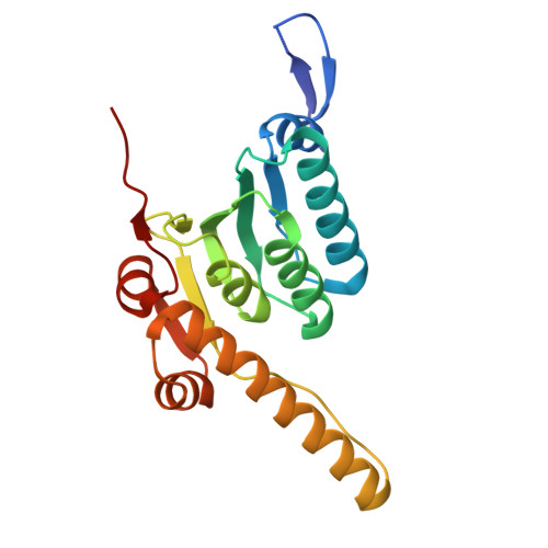 | |
UniProt | |||||
Entity Groups | |||||
| Sequence Clusters | 30% Identity50% Identity70% Identity90% Identity95% Identity100% Identity | ||||
| UniProt Group | Q2G036 | ||||
Sequence AnnotationsExpand | |||||
Reference Sequence | |||||
| Ligands 2 Unique | |||||
|---|---|---|---|---|---|
| ID | Chains | Name / Formula / InChI Key | 2D Diagram | 3D Interactions | |
| A1EDS Download:Ideal Coordinates CCD File | DA [auth L] GA [auth M] K [auth H] LA [auth N] P [auth I] | (12~{R})-7-[(4-chlorophenyl)methyl]-12-(ethoxymethyl)-11-[(3-fluorophenyl)methyl]-2,5,7,11-tetrazatricyclo[7.4.0.0^{2,6}]trideca-1(9),5-dien-8-one C26 H28 Cl F N4 O2 VCPJLNSMEZPNKY-JOCHJYFZSA-N |  | ||
| PEG (Subject of Investigation/LOI) Download:Ideal Coordinates CCD File | AA [auth L] BA [auth L] CA [auth L] EA [auth M] FA [auth M] | DI(HYDROXYETHYL)ETHER C4 H10 O3 MTHSVFCYNBDYFN-UHFFFAOYSA-N |  | ||
| Length ( Å ) | Angle ( ˚ ) |
|---|---|
| a = 123.64 | α = 90 |
| b = 123.64 | β = 90 |
| c = 107.07 | γ = 90 |
| Software Name | Purpose |
|---|---|
| PHENIX | refinement |
| Aimless | data scaling |
| XDS | data reduction |
| MOLREP | phasing |
| Funding Organization | Location | Grant Number |
|---|---|---|
| National Natural Science Foundation of China (NSFC) | China | 82473977 |


