Structural analysis of the ribosome assembly factor Nep1, an N1-specific pseudouridine methyltransferase, reveals mechanistic insights.
Saha, S., Kanaujia, S.P.(2025) FEBS J 292: 2338-2358
- PubMed: 39918246 Search on PubMed
- DOI: https://doi.org/10.1111/febs.70005
- Primary Citation Related Structures:
9JGB, 9JGC, 9JGD - PubMed Abstract:
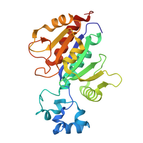
Nucleolar essential protein 1 (Nep1; also known as ribosomal RNA small subunit methyltransferase Nep1) is a crucial factor in forming small ribosomal subunits in eukaryotes and archaea. Nep1 possesses an S-adenosyl-L-methionine (SAM)-dependent SpoU-TrmD (SPOUT) ribosomal RNA (rRNA) methyltransferase (MTase) fold and catalyzes pseudouridine (Ψ) methylation at specific sites of the small subunit (SSU) rRNA. Mutations in Nep1 proteins result in a severe developmental disorder in humans and reduced growth in yeast, suggesting its role in ribosome biogenesis. In this study, the crystal structures of Nep1 from the archaebacterium Pyrococcus horikoshii (PhNep1), both in its apo and holo (adenosine or 5-methylthioadenosine bound) forms have been reported. The structural analysis of PhNep1 revealed an α/β fold featuring a deep trefoil knot akin to the SPOUT domain, with two novel extensions-a globular loop and a β-α-β extension. Moreover, the cofactor-binding site of PhNep1 exhibits a preformed pocket, topologically similar to that of other SPOUT-class MTases. Further, structural analysis of PhNep1 revealed that it forms a homodimer coordinated by inter-subunit hydrogen bonds and hydrophobic interactions. Moreover, the results of this study indicate that PhNep1 can specifically methylate consensus RNAs, having a pseudouridine (ψ) located at position 926 of helix 35 (h35) of 16S rRNA in P. horikoshii. The stability of the Nep1-RNA complex seems to be primarily assisted by the conserved arginine residues located at the dimeric interface.
- Department of Biosciences and Bioengineering, Indian Institute of Technology Guwahati, India.
Organizational Affiliation: