9JFM | pdb_00009jfm

Co-crystal Structure of sEH with tetrahydroberberine derivative


Experimental Data Snapshot

  • Method: X-RAY DIFFRACTION
  • Resolution: 3.13 Å
  • R-Value Free: 
    0.226 (Depositor), 0.227 (DCC) 
  • R-Value Work: 
    0.192 (Depositor), 0.193 (DCC) 
  • R-Value Observed: 
    0.194 (Depositor) 

Starting Model: experimental
View more details

wwPDB Validation   3D Report Full Report


Ligand Structure Quality Assessment 


This is version 1.1 of the entry. See complete history


Literature

Redesigning Berberines and Sanguinarines to Target Soluble Epoxide Hydrolase for Enhanced Anti-Inflammatory Efficacy.

Liu, X.Z.Du, X.Y.Xie, W.S.Ding, J.Zhu, M.Z.Feng, Z.Q.Wang, H.Feng, Y.Yu, M.J.Liu, S.M.Liu, W.T.Zhu, X.H.Liang, J.H.

(2024) J Med Chem 67: 22168-22190

  • DOI: https://doi.org/10.1021/acs.jmedchem.4c02202
  • Primary Citation Related Structures: 
    9JFM

  • PubMed Abstract: 

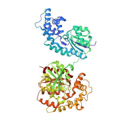
    Amino-berberine has remained underexplored due to limited biological evaluation and total synthesis approaches. In inflammation therapy, soluble Epoxide Hydrolase (sEH) is a promising target, yet natural scaffolds remain underutilized. Our study advances the field by redesigning natural compounds─berberine and sanguinarine─with strategic urea modifications and hydrogenated frameworks, creating novel sEH inhibitors with enhanced in vivo efficacy. Through total synthesis and structure-activity relationship studies of amino-berberine derivatives, chiral tetrahydroberberine ( R )-14i (coded LXZ-42 ) emerged as the most potent lead, with an IC 50 value of 1.20 nM. ( R )-14i showed reduced CYP enzyme impact, potent therapeutic effects on acute pancreatitis, no acute in vivo toxicity, and superior pharmacokinetic properties, with an oral bioavailability of 89.3%. Structural insights from crystallography of ( R )-14i bound to sEH revealed key interactions: three with the tetrahydroberberine framework and three hydrogen bonds with the urea group, highlighting ( R )-14i as a novel lead for sEH-targeted therapies in inflammation.


  • Organizational Affiliation
    • Key Laboratory of Medicinal Molecule Science and Pharmaceutical Engineering, School of Chemistry and Chemical Engineering, Beijing Institute of Technology, Beijing 102488, China.

Macromolecules
Find similar proteins by:  (by identity cutoff)  |  3D Structure
Entity ID: 1
MoleculeChains Sequence LengthOrganismDetailsImage
Bifunctional epoxide hydrolase 2555Homo sapiensMutation(s): 0 
Gene Names: EPHX2
EC: 3.3.2.10 (PDB Primary Data), 3.1.3.76 (PDB Primary Data)
UniProt & NIH Common Fund Data Resources
Find proteins for P34913 (Homo sapiens)
Explore P34913 
Go to UniProtKB:  P34913
PHAROS:  P34913
GTEx:  ENSG00000120915 
Entity Groups  
Sequence Clusters30% Identity50% Identity70% Identity90% Identity95% Identity100% Identity
UniProt GroupP34913
Sequence Annotations
Expand
  • Reference Sequence
Small Molecules
Experimental Data & Validation

Experimental Data

  • Method: X-RAY DIFFRACTION
  • Resolution: 3.13 Å
  • R-Value Free:  0.226 (Depositor), 0.227 (DCC) 
  • R-Value Work:  0.192 (Depositor), 0.193 (DCC) 
  • R-Value Observed: 0.194 (Depositor) 
Space Group: P 65 2 2
Unit Cell:
Length ( Å )Angle ( ˚ )
a = 91.89α = 90
b = 91.89β = 90
c = 244.96γ = 120
Software Package:
Software NamePurpose
PHENIXrefinement
xia2data reduction
xia2data scaling
PHENIXphasing

Structure Validation

View Full Validation Report


Ligand Structure Quality Assessment 


Entry History & Funding Information

Deposition Data


Funding OrganizationLocationGrant Number
Ministry of Science and Technology (MoST, China)China--

Revision History  (Full details and data files)

  • Version 1.0: 2025-09-10
    Type: Initial release
  • Version 1.1: 2026-04-01
    Changes: Database references