9JBD | pdb_00009jbd

Crystal Structure of the MoaE-like domain within Rv3323c from Mycobacterium tuberculosis


Experimental Data Snapshot

  • Method: X-RAY DIFFRACTION
  • Resolution: 2.40 Å
  • R-Value Free: 
    0.265 (Depositor), 0.263 (DCC) 
  • R-Value Work: 
    0.217 (Depositor), 0.218 (DCC) 
  • R-Value Observed: 
    0.222 (Depositor) 

Starting Model: experimental
View more details

wwPDB Validation   3D Report Full Report


This is version 1.0 of the entry. See complete history


Literature

Structural comparison of three MoaE proteins in Mycobacterium tuberculosis: Insights into molybdopterin synthase assembly and specificity.

Cho, H.J.Yang, S.H.Lee, H.S.Kang, B.S.

(2025) Biochem Biophys Res Commun 768: 151945-151945

  • DOI: https://doi.org/10.1016/j.bbrc.2025.151945
  • Primary Citation of Related Structures:  
    9JBD

  • PubMed Abstract: 

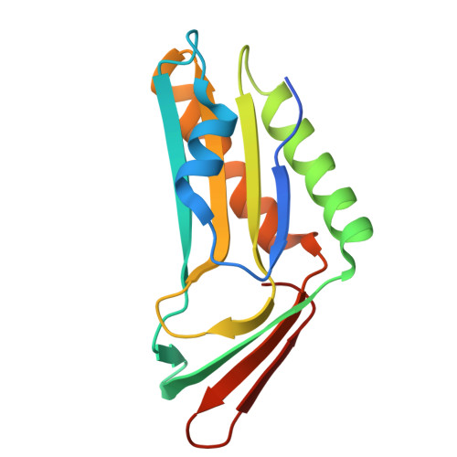
    Molybdoenzymes are essential for the survival and pathogenicity of Mycobacterium tuberculosis and require the molybdenum cofactor (MoCo). The biosynthesis of MoCo involves the molybdopterin (MPT) synthase complex, which is composed of the MoaD and MoaE subunits. The genome of M. tuberculosis encodes three homologs of MoaE: MoaE1, MoaE2, and MoaXE (the latter being a MoaE component of a MoaD-MoaE fusion protein known as MoaX), as well as three MoaD proteins. However, the structural basis for their functional specificity and interaction with MoaD partners remains unclear. We determined the crystal structures of all three MoaE proteins, revealing a conserved α/β hammerhead fold with distinct binding interface features resulting from minor sequence variations. Pull-down assays demonstrate that MoaE2 and MoaXE selectively interact with their cognate MoaD partners, while MoaE1 exhibits promiscuous binding to all MoaD forms. Although the structural plasticity of MoaE1 enables binding to three MoaD forms, it suggests that not all MoaE-MoaD combinations yield functional MPT synthase complexes, as structural rearrangements can lead to enzymatic inactivation. Our findings provide detailed insights into the molecular determinants that govern the assembly and specificity of MPT synthase in M. tuberculosis.


  • Organizational Affiliation
    • Department of Biological Sciences and Biotechnology, Chungbuk National University, Cheongju, 28644, Republic of Korea. Electronic address: hyojec@cbnu.ac.kr.

Macromolecules
Find similar proteins by:  (by identity cutoff)  |  3D Structure
Entity ID: 1
MoleculeChains Sequence LengthOrganismDetailsImage
Probable MoaD-MoaE fusion protein MoaX146Mycobacterium tuberculosis H37RvMutation(s): 0 
Gene Names: moaXRv3323c
EC: 2.8.1.12
UniProt
Find proteins for Q6MWY3 (Mycobacterium tuberculosis (strain ATCC 25618 / H37Rv))
Explore Q6MWY3 
Go to UniProtKB:  Q6MWY3
Entity Groups  
Sequence Clusters30% Identity50% Identity70% Identity90% Identity95% Identity100% Identity
UniProt GroupQ6MWY3
Sequence Annotations
Expand
  • Reference Sequence
Experimental Data & Validation

Experimental Data

  • Method: X-RAY DIFFRACTION
  • Resolution: 2.40 Å
  • R-Value Free:  0.265 (Depositor), 0.263 (DCC) 
  • R-Value Work:  0.217 (Depositor), 0.218 (DCC) 
  • R-Value Observed: 0.222 (Depositor) 
Space Group: P 65 2 2
Unit Cell:
Length ( Å )Angle ( ˚ )
a = 98.015α = 90
b = 98.015β = 90
c = 102.94γ = 120
Software Package:
Software NamePurpose
PHENIXrefinement
HKL-2000data scaling
HKL-2000data reduction
PHASERphasing

Structure Validation

View Full Validation Report


Entry History & Funding Information

Deposition Data


Funding OrganizationLocationGrant Number
National Research Foundation (NRF, Korea)Korea, Republic Of2021RIS-001
National Research Foundation (NRF, Korea)Korea, Republic Of2022R1F1A1074091

Revision History  (Full details and data files)

  • Version 1.0: 2025-08-27
    Type: Initial release