9J56 | pdb_00009j56

Functional Investigation of the SAM-Dependent Methyltransferases Rdmb in Anthracycline Biosynthesis


Experimental Data Snapshot

  • Method: X-RAY DIFFRACTION
  • Resolution: 2.10 Å
  • R-Value Free: 
    0.259 (Depositor), 0.263 (DCC) 
  • R-Value Work: 
    0.214 (Depositor), 0.228 (DCC) 

Starting Model: experimental
View more details

wwPDB Validation 3D Report Full Report

Validation slider image for 9J56

Ligand Structure Quality Assessment 


This is version 1.0 of the entry. See complete history

Literature

Functional investigation of the SAM-dependent methyltransferase RdmB in anthracycline biosynthesis.

Sang, M.Yang, Q.Guo, J.Feng, P.Ma, W.Zhang, W.

(2025) Synth Syst Biotechnol 10: 102-109

  • DOI: https://doi.org/10.1016/j.synbio.2024.09.002
  • Primary Citation Related Structures: 
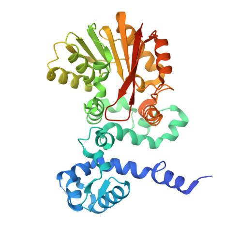
    9J56

  • PubMed Abstract: 

    A novel sub-class of S -adenosyl-l-methionine (SAM)-dependent methyltransferases catalyze atypical chemical transformations in the biosynthesis of anthracyclines. Exemplified by RdmB from Streptomyces purpurascens, it was found with 10-decarboxylative hydroxylation activity on anthracyclines. We herein investigated the catalytic activities of RdmB and discovered a previously unknown 4- O -methylation activity. The site-directed mutagenesis studies proved that the residue at position R307 and N260 are vital for the decarboxylative hydroxylation and 4- O -methylation, respectively, which define two distinct catalytic centers in RdmB. Furthermore, the multifunctionality of RdmB activity was found as cofactor-dependent and stepwise. Our findings expand the versatility and importance of methyltransferases and should aid studies to enrich the structural diversity and bioactivities of anthracyclines.


  • Organizational Affiliation
    • State Key Laboratory of Microbial Technology, Shandong University, Qingdao, Shandong, 266237, China.

Macromolecule Content 

  • Total Structure Weight: 81.47 kDa 
  • Atom Count: 5,270 
  • Modeled Residue Count: 669 
  • Deposited Residue Count: 748 
  • Unique protein chains: 1

Macromolecules

Find similar proteins by:|  3D Structure
Entity ID: 1
MoleculeChains  Sequence LengthOrganismDetailsImage
Aclacinomycin 10-hydroxylase RdmB
A, B
374Streptomyces purpurascensMutation(s): 0 
Gene Names: rdmB
EC: 4.1.1 (PDB Primary Data), 2.1.1 (UniProt)
UniProt
Find proteins for Q54527 (Streptomyces purpurascens)
Explore Q54527 
Go to UniProtKB:  Q54527
Entity Groups
Sequence Clusters30% Identity50% Identity70% Identity90% Identity95% Identity100% Identity
UniProt GroupQ54527
Sequence Annotations
Expand
Reference Sequence

Experimental Data & Validation

Experimental Data

  • Method: X-RAY DIFFRACTION
  • Resolution: 2.10 Å
  • R-Value Free:  0.259 (Depositor), 0.263 (DCC) 
  • R-Value Work:  0.214 (Depositor), 0.228 (DCC) 
Space Group: P 32 2 1
Unit Cell:
Length ( Å )Angle ( ˚ )
a = 79.997α = 90
b = 79.997β = 90
c = 234.203γ = 120
Software Package:
Software NamePurpose
PHENIXrefinement
HKL-3000data reduction
HKL-3000data scaling
PHENIXphasing

Structure Validation

View Full Validation Report


Ligand Structure Quality Assessment 


Entry History 

& Funding Information

Deposition Data


Funding OrganizationLocationGrant Number
National Natural Science Foundation of China (NSFC)China--

Revision History  (Full details and data files)

  • Version 1.0: 2024-11-06
    Type: Initial release