TPPP/p25 amyloid seeding activity as a specific biomarker for multiple system atrophy.
Zeng, S., Zhang, S., Zhang, S., Fan, Y., Xia, W., Chen, F., Huang, C., Lv, S., Lu, J., Sun, Y., Liu, K., Li, Y., Zhang, Y., Wang, J., Liu, C., Li, D.(2026) Cell
- PubMed: 42190663 Search on PubMed
- DOI: https://doi.org/10.1016/j.cell.2026.04.050
- Primary Citation Related Structures:
9J4D, 9J4E, 9J4F - PubMed Abstract:
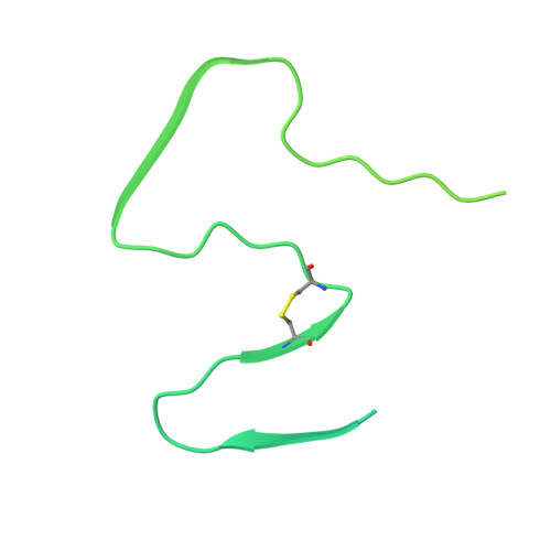
Detection of α-synuclein (α-syn) amyloid seeds in human biofluids has attracted great interest for clinical diagnosis of synucleinopathies. However, as a common biomarker, α-syn lacks specificity in reliably differentiating distinct disorders. Here, we report tubulin polymerization promoting protein (TPPP/p25) as a cerebrospinal fluid (CSF) biomarker for the specific diagnosis of multiple system atrophy (MSA). We demonstrate that native TPPP/p25 is self-protected against amyloid aggregation, while disease-related mutation disrupts this protection, triggering TPPP/p25 aggregation. Cryo-electron microscopy (cryo-EM) analysis reveals that the well-folded core domain (CORE) undergoes large conformational changes to mediate amyloid formation. Based on this insight, we developed a seed amplification assay using a minimized CORE (miniCORE) monomer, which detects TPPP/p25 amyloid seeds in CSF and robustly differentiates MSA from Parkinson's disease (PD) and other neurodegenerative diseases. Our findings establish misfolded TPPP/p25 as a promising, specific biomarker in biofluids for MSA diagnosis.
- Bio-X Institutes, Key Laboratory for the Genetics of Developmental and Neuropsychiatric Disorders (Ministry of Education), Shanghai Jiao Tong University, Shanghai 200030, China; Zhangjiang Institute for Advanced Study, Shanghai Jiao Tong University, Shanghai 200240, China.
Organizational Affiliation: