9J06 | pdb_00009j06

Cryo-EM structure of hOAT1 in complex with glibenclamide


Experimental Data Snapshot

  • Method: ELECTRON MICROSCOPY
  • Resolution: 3.68 Å
  • Aggregation State: PARTICLE 
  • Reconstruction Method: SINGLE PARTICLE 

wwPDB Validation   3D Report Full Report


This is version 1.1 of the entry. See complete history


Literature

Structures and membrane interactions of human OAT1 in complex with clinical used drugs.

Wu, X.Luo, Y.Feng, S.Ma, H.Ke, B.Wang, K.Su, Z.Yang, D.

(2025) Sci Adv 11: eads5405-eads5405

  • DOI: https://doi.org/10.1126/sciadv.ads5405
  • Primary Citation of Related Structures:  
    9J02, 9J04, 9J06

  • PubMed Abstract: 

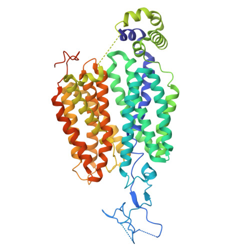
    Organic anion transporters (OATs) in mammals mediate the renal excretion of numerous structurally diverse organic anionic compounds. Therapeutically inhibiting OATs has emerged as a strategy to modulate the elimination or retention of these substrates. Among them, OAT1 plays a pivotal role in the pharmacokinetics and drug-drug interactions of a wide range of prescription medications. Despite extensive structural investigations, the molecular structure, and basis of polyspecific anionic drug recognition of human OAT1 (hOAT1) have remained elusive. Here, we present cryogenic electron microscopy structures of hOAT1 and its complexes with the antiviral drug cidofovir and an FDA-approved type II diabetes medication glibenclamide, respectively. Our findings reveal that both cidofovir and glibenclamide bind to a central binding site, capturing the transporter in inward-facing conformations. These structures elucidate how specific residues within the central site orchestrate the binding of chemically diverse inhibitors and provide a structural basis for the drug recognition mechanism of hOAT1.


  • Organizational Affiliation
    • Department of Urology, Institute of Urology (Laboratory of Reconstructive Urology), West China Hospital, Sichuan University, Chengdu, Sichuan 610044, China.

Macromolecules
Find similar proteins by:  (by identity cutoff)  |  3D Structure
Entity ID: 1
MoleculeChains Sequence LengthOrganismDetailsImage
Solute carrier family 22 member 6563Homo sapiensMutation(s): 0 
Gene Names: SLC22A6OAT1PAHT
Membrane Entity: Yes 
UniProt & NIH Common Fund Data Resources
Find proteins for Q4U2R8 (Homo sapiens)
Explore Q4U2R8 
Go to UniProtKB:  Q4U2R8
PHAROS:  Q4U2R8
GTEx:  ENSG00000197901 
Entity Groups  
Sequence Clusters30% Identity50% Identity70% Identity90% Identity95% Identity100% Identity
UniProt GroupQ4U2R8
Sequence Annotations
Expand
  • Reference Sequence
Small Molecules
Ligands 1 Unique
IDChains Name / Formula / InChI Key2D Diagram3D Interactions
GBM (Subject of Investigation/LOI)
Query on GBM

Download Ideal Coordinates CCD File 
B [auth A]5-chloro-N-(2-{4-[(cyclohexylcarbamoyl)sulfamoyl]phenyl}ethyl)-2-methoxybenzamide
C23 H28 Cl N3 O5 S
ZNNLBTZKUZBEKO-UHFFFAOYSA-N
Experimental Data & Validation

Experimental Data

  • Method: ELECTRON MICROSCOPY
  • Resolution: 3.68 Å
  • Aggregation State: PARTICLE 
  • Reconstruction Method: SINGLE PARTICLE 
EM Software:
TaskSoftware PackageVersion
MODEL REFINEMENTPHENIX

Structure Validation

View Full Validation Report


Entry History & Funding Information

Deposition Data


Funding OrganizationLocationGrant Number
National Natural Science Foundation of China (NSFC)China32222040
National Natural Science Foundation of China (NSFC)China32070049
Other governmentZYYC21006
Other government2022YFC2303700

Revision History  (Full details and data files)

  • Version 1.0: 2025-08-13
    Type: Initial release
  • Version 1.1: 2026-02-18
    Changes: Data collection, Database references