Vaspin identified as a DNA-binding serpin with functional consequences for protease inhibition.
Mohlis, K., Useini, A., Betat, H., Bonin, S., Broghammer, H., Nuwayhid, R., Langer, S., Morl, M., Strater, N., Heiker, J.T.(2026) FEBS J 293: 2119-2132
- PubMed: 40975878 Search on PubMed
- DOI: https://doi.org/10.1111/febs.70270
- Primary Citation Related Structures:
9IH0 - PubMed Abstract:
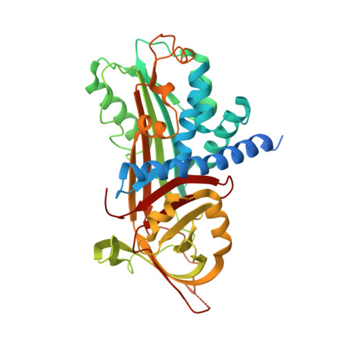
Vaspin is highly expressed not only in the skin but also in the liver and adipose tissue. It counteracts inflammation and oxidative stress in inflammatory skin diseases, obesity, and associated metabolic disorders, in part by inhibiting the kallikrein proteases KLK7 and KLK14. Vaspin binds the cell-surface low-density lipoprotein receptor-related protein 1 (LRP1) with nanomolar affinity, and is rapidly internalized into adipocytes and other cells. We found intracellular vaspin partially localized in the nucleus. Since vaspin binds heparin and inorganic polyphosphates, we investigated the DNA binding of vaspin. Using DNA-affinity chromatography and differential radial capillary action of ligand assays, we found high-affinity binding to random sequences of single- and double-stranded DNA for both vaspin and KLK7. Furthermore, KLK7 inhibition was accelerated fivefold in the presence of DNA molecules at least 40 bases in length. We previously identified the heparin-binding site at a basic patch on the central beta-sheet A of vaspin. In the current work, we determined the crystal structure of polyphosphate P45-bound vaspin, which confirmed previously identified residues mutated to generate a nonheparin-binding (NHB) vaspin variant. While NHB vaspin failed to bind heparin and polyP45, it still bound DNA with high affinity and accelerated protease inhibition. Mutation of closely spaced basic residues in helix A and helix G did not significantly alter DNA binding. In conclusion, we have identified vaspin as the second human DNA-binding serpin. While the exact mode of the nonspecific interaction remains unclear, it accelerates protease inhibition and likely contributes to the nuclear localization observed for internalized vaspin and may allow for intracellular effects.
- Helmholtz Institute for Metabolic, Obesity and Vascular Research (HI-MAG) of the Helmholtz Zentrum München at the University of Leipzig and University Hospital Leipzig, Germany.
Organizational Affiliation: