9IF4 | pdb_00009if4

Structure of the Mycobacterium Tuberculosis ClpC1P1P2 complex bound to the activator Bz-Leu-Leu


Experimental Data Snapshot

  • Method: ELECTRON MICROSCOPY
  • Resolution: 3.09 Å
  • Aggregation State: PARTICLE 
  • Reconstruction Method: SINGLE PARTICLE 

wwPDB Validation 3D Report Full Report

Validation slider image for 9IF4

This is version 1.1 of the entry. See complete history

Literature

Activation mechanism and structural assembly of the Mycobacterium tuberculosis ClpP1P2 protease and its associated ATPases.

Weinhaupl, K.Akopian, T.Semchonok, D.A.Krandor, O.Arranz, R.Bueno-Carrasco, M.T.Gragera, M.Chevreuil, M.Raynal, B.Zinga, S.Liu, Y.Lai, J.Wu, W.Bachovchin, W.Gauto, D.Goldberg, A.Rubin, E.Fraga, H.

(2026) Cell Rep 45: 117400-117400

  • DOI: https://doi.org/10.1016/j.celrep.2026.117400
  • Primary Citation Related Structures: 
    9IF4

  • PubMed Abstract: 

    Supramolecular assemblies are fundamental to cellular biochemical processes, relying on their dynamic nature to perform essential functions. The protease ClpP1P2, in association with ATPase partners ClpC1 or ClpX, is critical for the survival of Mycobacterium tuberculosis (Mtb). While the ClpP1P2 complex requires activation by specific N-blocked dipeptides to exhibit proteolytic activity in vitro, the mechanism of in vivo activation remains unclear. In this study, we use cryo-electron microscopy (cryo-EM) to determine the structure of the ClpC1P1P2 complex, revealing a highly asymmetric architecture with ClpC1 bound to the ClpP1P2 protease barrel. The activator dipeptide is observed only in the ClpP2 active site, while the ClpP1 entry pore remains closed. Molecular crowding agents promote the formation of larger ClpXP1P2 and ClpC1P1P2 complexes, enhancing structural stability and enzymatic activity. These findings suggest that molecular crowding stabilizes these complexes and promotes their activation, providing new insights into ClpC1P1P2 structural dynamics and function.


  • Organizational Affiliation
    • i3S, Instituto de Investigação e Inovação em Saúde, Universidade do Porto, Porto, Portugal.

Macromolecule Content 

  • Total Structure Weight: 741.77 kDa 
  • Atom Count: 45,182 
  • Modeled Residue Count: 5,762 
  • Deposited Residue Count: 6,641 
  • Unique protein chains: 5

Macromolecules

Find similar proteins by:|  3D Structure
Entity ID: 1
MoleculeChains  Sequence LengthOrganismDetailsImage
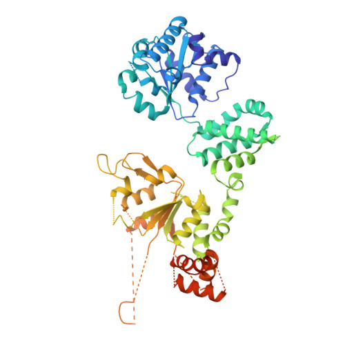
ATP-dependent Clp protease ATP-binding subunit ClpC1
A, B, C, D, E
A, B, C, D, E, F
658Mycobacterium tuberculosisMutation(s): 0 
Gene Names: clpC1Rv3596cMTCY07H7B.26
UniProt
Find proteins for P9WPC9 (Mycobacterium tuberculosis (strain ATCC 25618 / H37Rv))
Explore P9WPC9 
Go to UniProtKB:  P9WPC9
Entity Groups
Sequence Clusters30% Identity50% Identity70% Identity90% Identity95% Identity100% Identity
UniProt GroupP9WPC9
Sequence Annotations
Expand
Reference Sequence
Find similar proteins by:|  3D Structure
Entity ID: 2
MoleculeChains  Sequence LengthOrganismDetailsImage
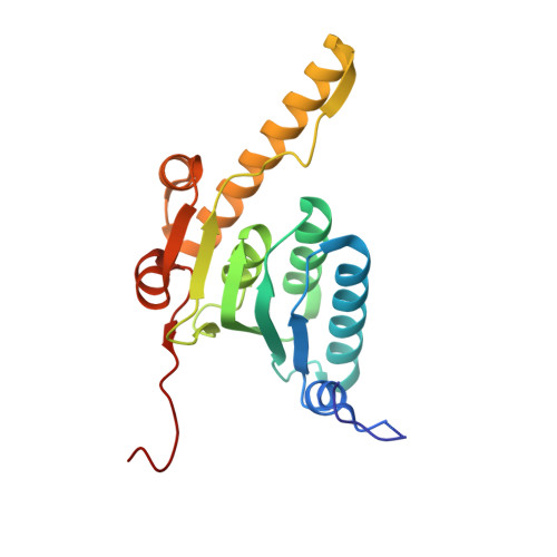
ATP-dependent Clp protease proteolytic subunit 2200Mycobacterium tuberculosisMutation(s): 0 
Gene Names: clpP2Rv2460cMTV008.16c
EC: 3.4.21.92
UniProt
Find proteins for P9WPC3 (Mycobacterium tuberculosis (strain ATCC 25618 / H37Rv))
Explore P9WPC3 
Go to UniProtKB:  P9WPC3
Entity Groups
Sequence Clusters30% Identity50% Identity70% Identity90% Identity95% Identity100% Identity
UniProt GroupP9WPC3
Sequence Annotations
Expand
Reference Sequence
Find similar proteins by:|  3D Structure
Entity ID: 3
MoleculeChains  Sequence LengthOrganismDetailsImage
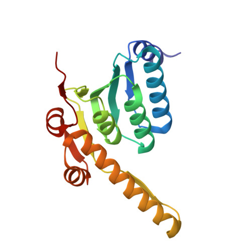
ATP-dependent Clp protease proteolytic subunit 1179Mycobacterium tuberculosisMutation(s): 0 
Gene Names: clpP1clpPRv2461cMTV008.17c
EC: 3.4.21.92
UniProt
Find proteins for P9WPC5 (Mycobacterium tuberculosis (strain ATCC 25618 / H37Rv))
Explore P9WPC5 
Go to UniProtKB:  P9WPC5
Entity Groups
Sequence Clusters30% Identity50% Identity70% Identity90% Identity95% Identity100% Identity
UniProt GroupP9WPC5
Sequence Annotations
Expand
Reference Sequence
Find similar proteins by:  Sequence   |   3D Structure  
Entity ID: 4
MoleculeChains  Sequence LengthOrganismDetailsImage
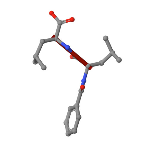
activator Bz-Leu-Leu2synthetic constructMutation(s): 0 
Sequence Annotations
Expand
Reference Sequence
Find similar proteins by:|  3D Structure
Entity ID: 5
MoleculeChains  Sequence LengthOrganismDetailsImage
Unknown peptideBA [auth X]26Mycobacterium tuberculosisMutation(s): 0 
Sequence Annotations
Expand
Reference Sequence

Small Molecules

Ligands 2 Unique
IDChains Name / Formula / InChI Key2D Diagram3D Interactions
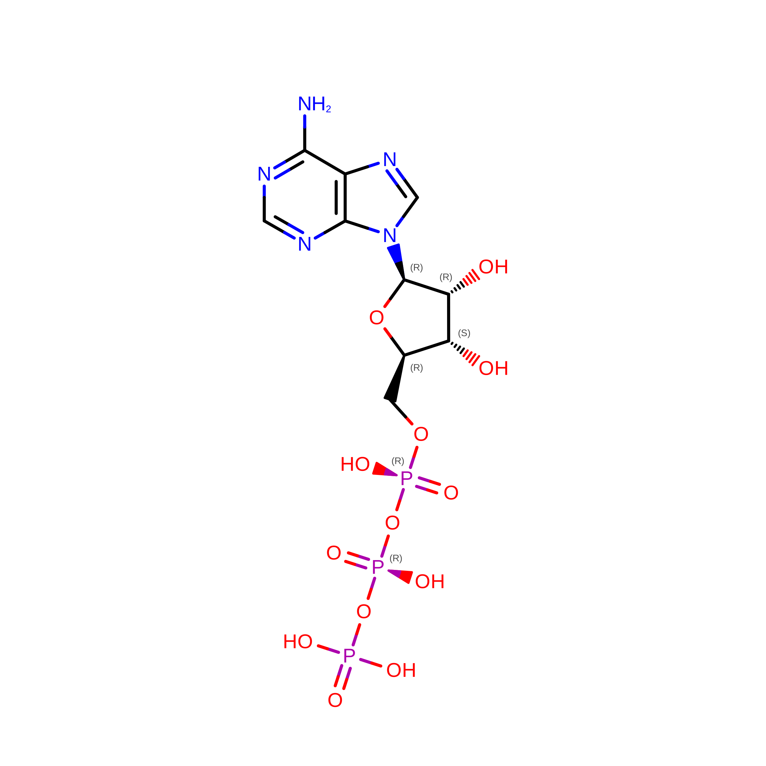
ATP
(Subject of Investigation/LOI)

Query on ATP


Download:Ideal Coordinates CCD File
EA [auth B]
FA [auth B]
GA [auth C]
HA [auth C]
IA [auth D]
EA [auth B],
FA [auth B],
GA [auth C],
HA [auth C],
IA [auth D],
JA [auth D],
KA [auth E],
LA [auth E]
ADENOSINE-5'-TRIPHOSPHATE
C10 H16 N5 O13 P3
ZKHQWZAMYRWXGA-KQYNXXCUSA-N
ADP
(Subject of Investigation/LOI)

Query on ADP


Download:Ideal Coordinates CCD File
CA [auth A],
DA [auth A],
MA [auth F],
NA [auth F]
ADENOSINE-5'-DIPHOSPHATE
C10 H15 N5 O10 P2
XTWYTFMLZFPYCI-KQYNXXCUSA-N
Modified Residues  2 Unique
IDChains TypeFormula2D DiagramParent
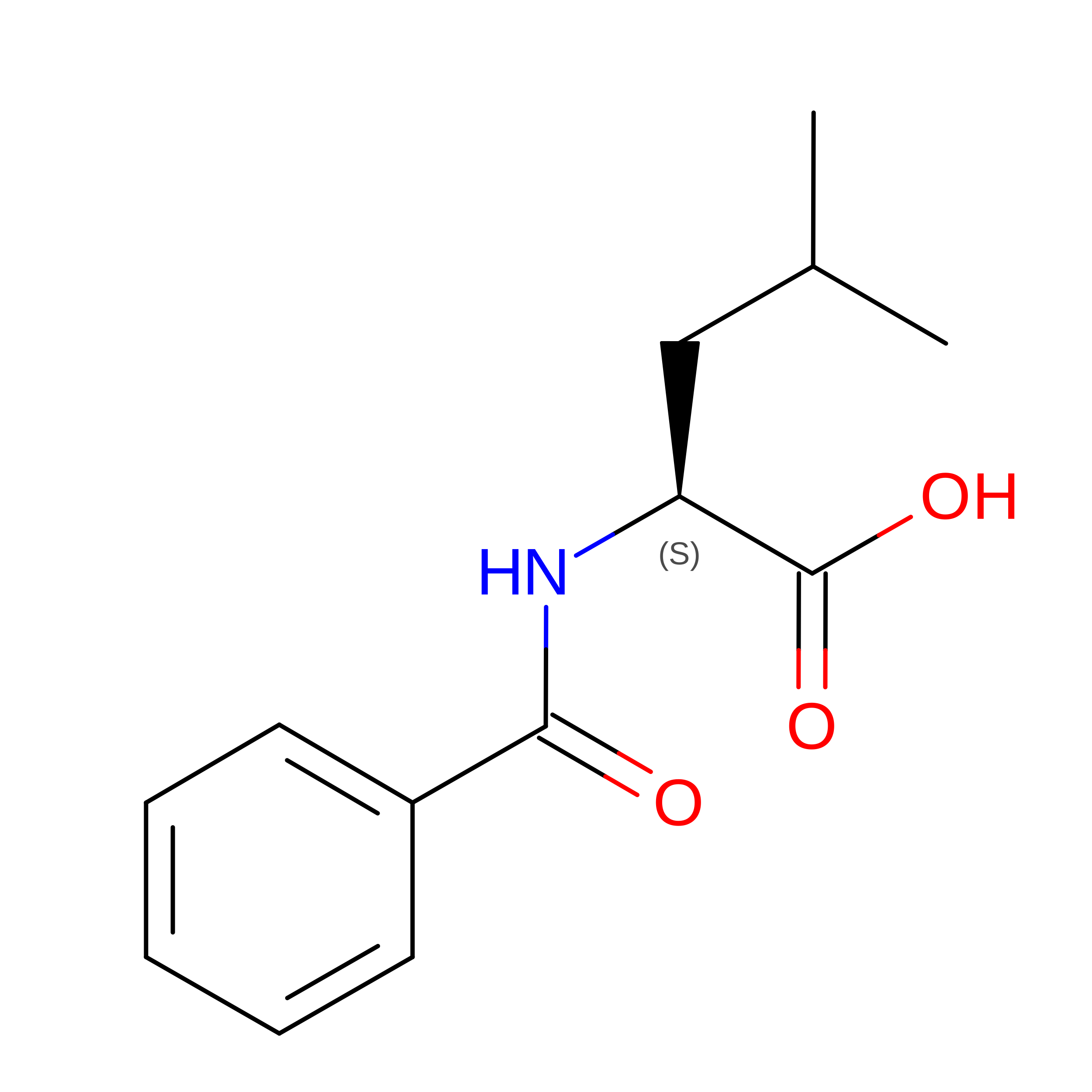
S0R
Query on S0R
AA [auth b]
U
V
W
X [auth Y]
AA [auth b],
U,
V,
W,
X [auth Y],
Y [auth Z],
Z [auth a]
L-PEPTIDE LINKINGC13 H17 N O3LEU
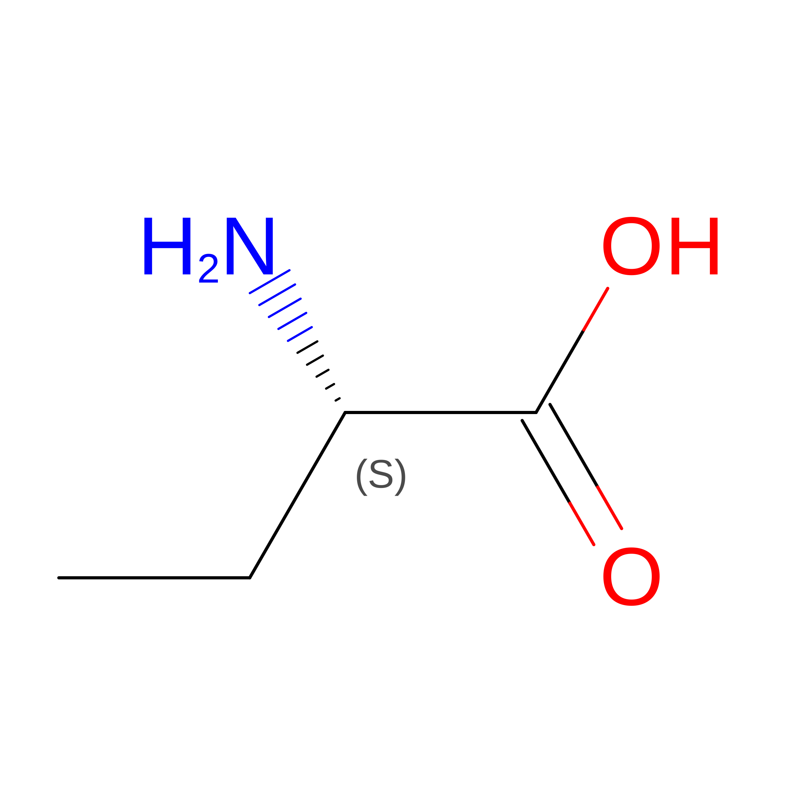
UNK
Query on UNK
BA [auth X]L-PEPTIDE LINKINGC4 H9 N O2

--

Experimental Data & Validation

Experimental Data

  • Method: ELECTRON MICROSCOPY
  • Resolution: 3.09 Å
  • Aggregation State: PARTICLE 
  • Reconstruction Method: SINGLE PARTICLE 
EM Software:
TaskSoftware PackageVersion
MODEL REFINEMENTPHENIX1.21.1_5286
RECONSTRUCTIONcryoSPARC

Structure Validation

View Full Validation Report


Entry History 

& Funding Information

Deposition Data


Funding OrganizationLocationGrant Number
Other governmentInstruct-ERIC 30641, 26164

Revision History  (Full details and data files)

  • Version 1.0: 2025-08-27
    Type: Initial release
  • Version 1.1: 2026-09-09
    Changes: Data collection, Database references