Molecular basis of antibiotic sensing by the TetR family regulator CecR - a structural perspective.
Pietrzyk-Brzezinska, A.J., Koczurowska, A., Orlikowska, M., Nielipinski, M., Nielipinska, D., Sekula, B.(2026) FEBS J 293: 1801-1817
- PubMed: 41194554 Search on PubMed
- DOI: https://doi.org/10.1111/febs.70318
- Primary Citation Related Structures:
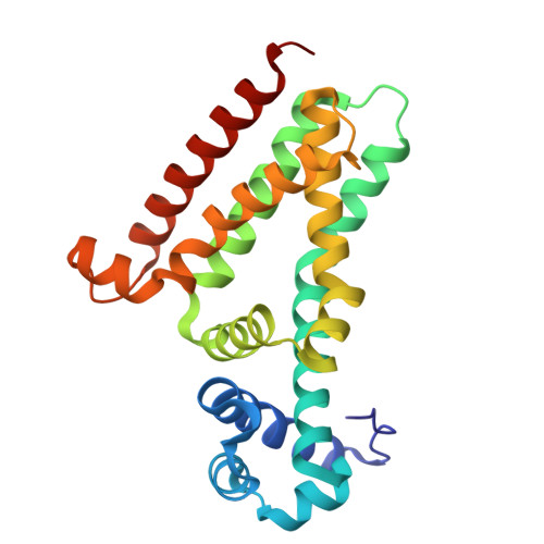
9I1X - PubMed Abstract:
Escherichia coli HTH-type transcriptional dual regulator CecR belongs to TetR family regulators (TFRs), which regulate the expression of genes enabling bacteria to survive under stress conditions. Previous studies (Yamanaka et al., Microbiology 2016; 162: 1253-1264) showed that CecR senses the presence of antibiotics, cephalosporins and chloramphenicol, in the cell and activates the expression of a putative drug efflux pump. Although CecR is present in many pathogenic strains of Escherichia and Salmonella genera, this regulator is poorly characterized. Here, we report the first crystal structure of E. coli CecR. Each protomer of the CecR homodimer is composed of an N-terminal DNA-binding and a C-terminal ligand-binding domain. In addition to nine canonical TetR α-helices, CecR contains structural elements characteristic of TetR subfamily D. The ligand-binding cavity of CecR has a tunnel-like shape, not common in TFRs. Unexpectedly, the CecR-ligand-binding cavity contained polyethylene glycol (PEG) fragments, originating from crystallization solution, and suggesting a potential site for effector binding. Additionally, the affinity of CecR to various antibiotics was determined. The strongest interactions were observed for CecR and cefepime, a representative of the fourth-generation cephalosporins. Molecular docking of the analyzed antibiotics into the ligand-binding tunnel of CecR indicated the amino acid residues important for ligand recognition. The CecR structure reported here provides the first structural information on the ligand-binding cavity and ligand recognition by CecR. As CecR is an important regulator, widespread among pathogenic bacteria belonging to the Enterobacteriales order, the results of our study are an important contribution to the understanding of the CecR-related mechanisms underlying antimicrobial resistance.
- Institute of Molecular and Industrial Biotechnology, Faculty of Biotechnology and Food Sciences, Lodz University of Technology, Lodz, Poland.
Organizational Affiliation: