Convergent evolution of distinct D-ribulose utilisation pathways in attaching and effacing pathogens.
Cottam, C., Bowran, K., White, R.T., Basle, A., Josts, I., Connolly, J.P.R.(2025) Nat Commun 16: 6976-6976
- PubMed: 40730545 Search on PubMedSearch on PubMed Central
- DOI: https://doi.org/10.1038/s41467-025-62476-5
- Primary Citation Related Structures:
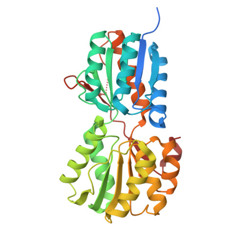
9I1M - PubMed Abstract:
Attaching and effacing pathogens overcome colonisation resistance by competing with metabolically similar organisms for limited resources. Enterohaemorrhagic E. coli (EHEC) utilises the pathogenicity island-encoded Accessory ʟ-arabinose Uptake (Aau) transporter to effectively colonise the mouse gut, hypothesised to be achieved via an enhanced capacity to scavenge ʟ-arabinose. Aau is regulated exclusively in response to ʟ-arabinose, but it is unclear how this system specifically benefits EHEC in vivo. Here, we show that Aau displays a > 200-fold higher affinity for the monosaccharide D-ribulose, over ʟ-arabinose. EHEC cannot grow on D-ribulose as a sole carbon source and this sugar does not trigger aau transcription. However, Aau effectively transports D-ribulose into the cell only in the presence of ʟ-arabinose, where it feeds into the pentose phosphate pathway, after phosphorylation by the ʟ-ribulokinase AraB, thus providing EHEC a significant fitness advantage. EHEC has therefore evolved a mechanism of hijacking the canonical ʟ-arabinose utilisation machinery to promote D-ribulose utilisation in vivo. Furthermore, Citrobacter rodentium encodes an analogous system that exclusively transports D-ribulose and metabolises it via a dedicated D-ribulokinase. These unique mechanisms of D-ribulose utilisation suggest that convergent evolution has driven the ability of distinct pathogenic species to exploit this nutrient during invasion of the gut niche.
- Newcastle University Biosciences Institute, Newcastle University, Newcastle-upon-Tyne, UK.
Organizational Affiliation: