9HYE | pdb_00009hye

Structure of the Mus musclus Langerin carbohydrate recognition domain with depleted Calcium


Experimental Data Snapshot

  • Method: X-RAY DIFFRACTION
  • Resolution: 1.64 Å
  • R-Value Free: 
    0.215 (Depositor), 0.214 (DCC) 
  • R-Value Work: 
    0.154 (Depositor), 0.153 (DCC) 
  • R-Value Observed: 
    0.157 (Depositor) 

Starting Model: experimental
View more details

wwPDB Validation   3D Report Full Report


This is version 1.2 of the entry. See complete history


Literature

Calcium Competitive Inhibition of Langerin by Thiazolopyrimidinones.

Ning, Y.Efrem, N.L.Amoussa, M.Turhan, E.Zheng, D.Lefebre, J.Ruwolt, M.Neu, U.Besch, M.Loll, B.Kurzbach, D.Kohnke, J.Nazare, M.Rademacher, C.

(2025) J Med Chem 68: 24924-24934

  • DOI: https://doi.org/10.1021/acs.jmedchem.5c01756
  • Primary Citation of Related Structures:  
    9HYE

  • PubMed Abstract: 

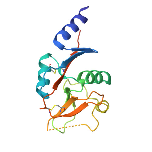
    C-Type lectins are a large family of carbohydrate-binding proteins. Langerin is a member of this family and is expressed by Langerhans cells, involved in pathogen recognition and innate immune activation, making it a target for small-molecule modulation in immunology and infectious diseases. We previously identified thiazolopyrimidinones as a series of allosteric inhibitors, but the underlying mechanism remained unclear. In this study, 43 Ca NMR demonstrated that these fragments induce Ca 2+ release from the receptor. Our ITC data suggested a competitive relationship between inhibitors and Ca 2+ , which was further validated by 19 F NMR spectroscopy showing inhibition of carbohydrate binding. Surprisingly, the fragment binding site was found to be located beneath the long loop, which supports the dynamic nature of the long loop being highly Ca 2+ dependent. Our findings provide insight into the novel Ca 2+ -competitive inhibitory mechanism of murine langerin and are the first report on such an inhibitory mechanism for a C-type lectin.


  • Organizational Affiliation
    • Department of Pharmaceutical Sciences, University of Vienna, Josef-Holaubek-Platz 2, Vienna 1090, Austria.

Macromolecules
Find similar proteins by:  (by identity cutoff)  |  3D Structure
Entity ID: 1
MoleculeChains Sequence LengthOrganismDetailsImage
C-type lectin domain family 4 member K
A, B
154Mus musculusMutation(s): 1 
Gene Names: Cd207Clec4k
UniProt
Find proteins for Q8VBX4 (Mus musculus)
Explore Q8VBX4 
Go to UniProtKB:  Q8VBX4
Entity Groups  
Sequence Clusters30% Identity50% Identity70% Identity90% Identity95% Identity100% Identity
UniProt GroupQ8VBX4
Sequence Annotations
Expand
  • Reference Sequence
Experimental Data & Validation

Experimental Data

  • Method: X-RAY DIFFRACTION
  • Resolution: 1.64 Å
  • R-Value Free:  0.215 (Depositor), 0.214 (DCC) 
  • R-Value Work:  0.154 (Depositor), 0.153 (DCC) 
  • R-Value Observed: 0.157 (Depositor) 
Space Group: P 1 21 1
Unit Cell:
Length ( Å )Angle ( ˚ )
a = 34.917α = 90
b = 80.706β = 105.25
c = 52.179γ = 90
Software Package:
Software NamePurpose
PHENIXrefinement
XDSdata reduction
XSCALEdata scaling
PHASERphasing

Structure Validation

View Full Validation Report


Entry History & Funding Information

Deposition Data


Funding OrganizationLocationGrant Number
German Research Foundation (DFG)Germany--

Revision History  (Full details and data files)

  • Version 1.0: 2025-11-19
    Type: Initial release
  • Version 1.1: 2025-11-26
    Changes: Database references
  • Version 1.2: 2025-12-24
    Changes: Database references