9HY1 | pdb_00009hy1

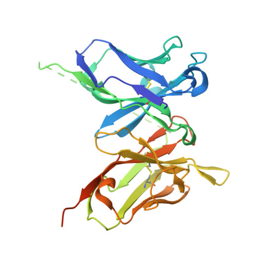
sc-4E (scFv derived from the mAb 4E1 against CD93) crystallized at pH 7.5 (dimeric state from gel filtration)


Experimental Data Snapshot

  • Method: X-RAY DIFFRACTION
  • Resolution: 1.81 Å
  • R-Value Free: 
    0.196 (Depositor), 0.196 (DCC) 
  • R-Value Work: 
    0.169 (Depositor), 0.170 (DCC) 
  • R-Value Observed: 
    0.170 (Depositor) 

Starting Model: experimental
View more details

wwPDB Validation 3D Report Full Report

Validation slider image for 9HY1

This is version 1.0 of the entry. See complete history. 

Literature

Structural and antigen-binding surface definition of an anti-CD93 monoclonal antibody for the treatment of degenerative vascular eye diseases.

Raucci, L., Perrone, C.D., Barbera, S., de Boer, L.J., Tosi, G.M., Brunetti, J., Bracci, L., Pozzi, C., Galvagni, F., Orlandini, M.

(2025) Int J Biol Macromol 309: 143118-143118

  • DOI: https://doi.org/10.1016/j.ijbiomac.2025.143118
  • Primary Citation Related Structures: 
    9HY1, 9HY2, 9HY3, 9HY5

  • PubMed Abstract: 

    CD93 is a receptor predominantly expressed on the surface of endothelial cells, where it plays a pivotal role in angiogenesis through its interaction with the extracellular matrix. In our previous studies, we identified the monoclonal antibody 4E1 as a potent inhibitor of angiogenesis by targeting the CD93-Multimerin-2 axis. Here, we report the development of 4E1 as a recombinant whole immunoglobulin and a single-chain variable fragment, designated sc-4E. Both formats retained the binding properties of the parental monoclonal antibody and exhibited comparable inhibitory effects on endothelial cell migration and differentiation. To elucidate the molecular basis of the 4E1-CD93 interaction, we initially employed machine learning-based modeling and docking analyses of the variable heavy and light domains of 4E1. Subsequent crystallographic analysis of sc-4E provided high-resolution structural insights, confirming and validating the predicted model. Further docking experiments and molecular dynamics simulations using the crystallographic structures of CD93 and sc-4E revealed that the interaction is primarily mediated by the CDR-H3 and CDR-L2 loops. Notably, these regions engage with the sushi-like domain of CD93, which is critical for its interaction with Multimerin-2. This comprehensive structural and functional characterization of 4E1 and sc-4E underscores their potential as anti-angiogenic agents. By effectively inhibiting endothelial cell migration and differentiation, 4E1 derivatives represent promising therapeutic candidates for the treatment of ocular vascular diseases driven by pathological angiogenesis.


  • Organizational Affiliation: 
    • Department of Biotechnology, Chemistry and Pharmacy, University of Siena, Siena 53100, Italy.

Macromolecule Content 

  • Total Structure Weight: 29.22 kDa 
  • Atom Count: 2,097 
  • Modeled Residue Count: 232 
  • Deposited Residue Count: 269 
  • Unique protein chains: 1

Macromolecules

Find similar proteins by:|  3D Structure
Entity ID: 1
MoleculeChains  Sequence LengthOrganismDetailsImage
sc-4E (scFv derived from the mAb 4E1 against CD93)269Mus musculusMutation(s): 0 
Entity Groups
Sequence Clusters30% Identity50% Identity70% Identity90% Identity95% Identity100% Identity
Sequence Annotations
Expand
Reference Sequence

Small Molecules

Ligands 4 Unique
IDChains Name / Formula / InChI Key2D Diagram3D Interactions
IMD

Query on IMD


Download:Ideal Coordinates CCD File
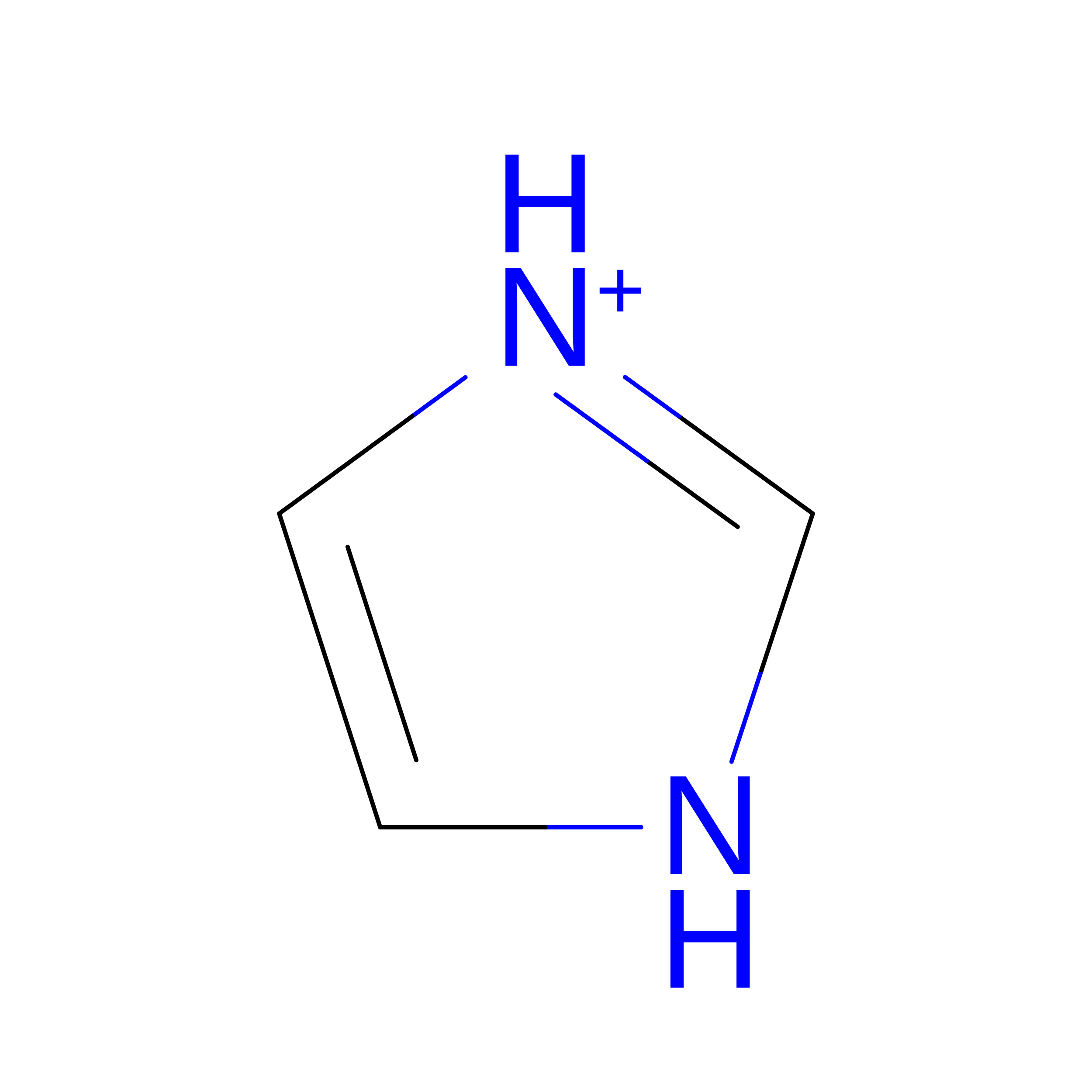
E [auth A]IMIDAZOLE
C3 H5 N2
RAXXELZNTBOGNW-UHFFFAOYSA-O
EDO

Query on EDO


Download:Ideal Coordinates CCD File
C [auth A],
D [auth A],
F [auth A]
1,2-ETHANEDIOL
C2 H6 O2
LYCAIKOWRPUZTN-UHFFFAOYSA-N
CL

Query on CL


Download:Ideal Coordinates CCD File
B [auth A],
I [auth A]
CHLORIDE ION
Cl
VEXZGXHMUGYJMC-UHFFFAOYSA-M
NA
(Subject of Investigation/LOI)

Query on NA


Download:Ideal Coordinates CCD File
G [auth A],
H [auth A]
SODIUM ION
Na
FKNQFGJONOIPTF-UHFFFAOYSA-N

Experimental Data & Validation

Experimental Data

  • Method: X-RAY DIFFRACTION
  • Resolution: 1.81 Å
  • R-Value Free:  0.196 (Depositor), 0.196 (DCC) 
  • R-Value Work:  0.169 (Depositor), 0.170 (DCC) 
  • R-Value Observed: 0.170 (Depositor) 
Space Group: P 32 2 1
Unit Cell:
Length ( Å )Angle ( ˚ )
a = 64.369α = 90
b = 64.369β = 90
c = 159.88γ = 120
Software Package:
Software NamePurpose
REFMACrefinement
XDSdata reduction
Aimlessdata scaling
MOLREPphasing

Structure Validation

View Full Validation Report


Entry History 

& Funding Information

Deposition Data


Funding OrganizationLocationGrant Number
European Union (EU)European UnionBC63C22000680007

Revision History  (Full details and data files)

  • Version 1.0: 2025-04-30
    Type: Initial release