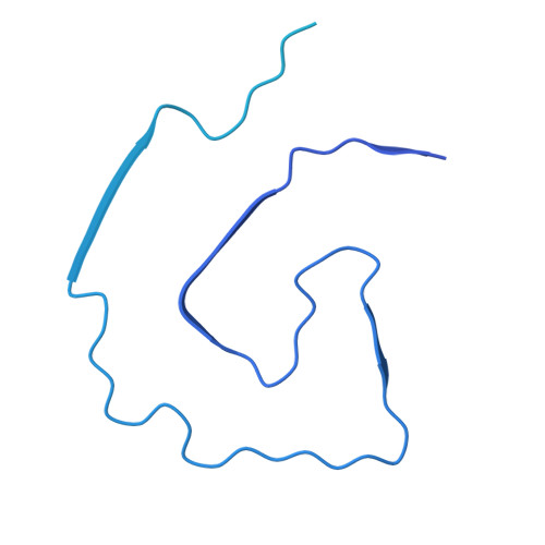
9HX4 | pdb_00009hx4

Amyloid fibril of apolipoprotein A-IV


Experimental Data Snapshot

  • Method: ELECTRON MICROSCOPY
  • Resolution: 3.30 Å
  • Aggregation State: HELICAL ARRAY 
  • Reconstruction Method: HELICAL 

wwPDB Validation 3D Report Full Report

Validation slider image for 9HX4

This is version 1.0 of the entry. See complete history. 

Literature

Apolipoprotein A-IV fibrils: structural diagnosis of mixed cardiac amyloidosis.

Aibara, S., Kassner, A., Wong, E., Klingel, K., Papworth, M., Althage, M., Wang, Q.D., Correia, C., Milting, H., de Oliveira, T.M.

(2025) Nat Commun 16: 9276-9276

  • DOI: https://doi.org/10.1038/s41467-025-64902-0
  • Primary Citation Related Structures: 
    9HX3, 9HX4

  • PubMed Abstract: 

    Cardiac amyloidosis (CA) occurs when misfolded proteins deposit as fibrils in the extracellular space of the heart. The fibrillogenic properties of apolipoprotein A-IV (ApoAIV) have been histologically observed and associated with CA pathogenesis. We report the structure of an ApoAIV amyloid from a patient's heart, which coexist amongst transthyretin (TTR) amyloids. These cases of undetected mixed CA highlight the importance of developing broad-spectrum anti-amyloid treatments to improve outcomes in patients.


  • Organizational Affiliation: 
    • Protein Sciences, Structure, and Biophysics, Discovery Sciences, R&D, AstraZeneca, Cambridge, UK.

Macromolecule Content 

  • Total Structure Weight: 454.34 kDa 
  • Atom Count: 4,840 
  • Modeled Residue Count: 610 
  • Deposited Residue Count: 3,960 
  • Unique protein chains: 1

Macromolecules

Find similar proteins by:|  3D Structure
Entity ID: 1
MoleculeChains  Sequence LengthOrganismDetailsImage
Apolipoprotein A-IV396Homo sapiensMutation(s): 0 
UniProt & NIH Common Fund Data Resources
Find proteins for P06727 (Homo sapiens)
Explore P06727 
Go to UniProtKB:  P06727
PHAROS:  P06727
GTEx:  ENSG00000110244 
Entity Groups
Sequence Clusters30% Identity50% Identity70% Identity90% Identity95% Identity100% Identity
UniProt GroupP06727
Sequence Annotations
Expand
Reference Sequence

Experimental Data & Validation

Experimental Data

  • Method: ELECTRON MICROSCOPY
  • Resolution: 3.30 Å
  • Aggregation State: HELICAL ARRAY 
  • Reconstruction Method: HELICAL 
EM Software:
TaskSoftware PackageVersion
MODEL REFINEMENTPHENIX1.21.1_5286:

Structure Validation

View Full Validation Report


Entry History 

& Funding Information

Deposition Data


Funding OrganizationLocationGrant Number
Not funded--

Revision History  (Full details and data files)

  • Version 1.0: 2025-11-05
    Type: Initial release