9HVI | pdb_00009hvi

PSMA in complex with nanobody 8


Experimental Data Snapshot

  • Method: ELECTRON MICROSCOPY
  • Resolution: 2.46 Å
  • Aggregation State: PARTICLE 
  • Reconstruction Method: SINGLE PARTICLE 

wwPDB Validation   3D Report Full Report


This is version 1.1 of the entry. See complete history


Literature

Structural analysis of nanobody interactions with their prostate-specific membrane antigen binding epitopes.

Alon-Zchut, G.Zalk, R.Huynh, T.T.Zalutsky, M.R.Weizmann, Y.Zarivach, R.Papo, N.

(2025) Int J Biol Macromol 320: 145693-145693

  • DOI: https://doi.org/10.1016/j.ijbiomac.2025.145693
  • Primary Citation of Related Structures:  
    9HLW, 9HVI, 9HVK, 9HVL

  • PubMed Abstract: 

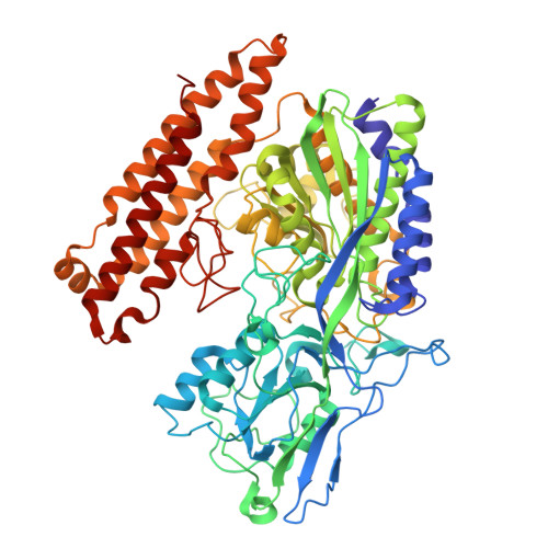
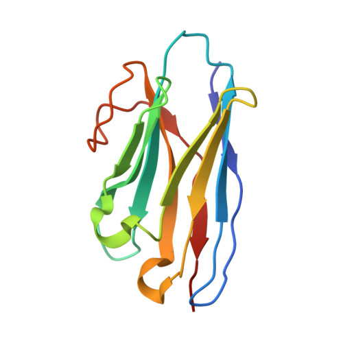
    Prostate-specific membrane antigen (PSMA), overexpressed in prostate cancer, is a promising target for diagnostics and therapy. However, the monoclonal antibodies in current use for PSMA targeting and inhibition have suboptimal activities due to their poor tissue and cell penetration and slow normal tissue clearance. Potentially superior alternatives are nanobodies (NBs), the single-chain variable domains of heavy-chain antibodies derived from camelids. The advantages of NBs include small size (~15 kDa), ability to bind hidden epitopes, and rapid clearance. In contrast to most known PSMA inhibitors, which bind to the same catalytic site in PMSA, NBs can bind to different PSMA epitopes, facilitating heterovalent binding strategies that could enhance their therapeutic and diagnostic potential. The objective of this study was to map these binding epitopes and hence to acquire an atomic-resolution understanding of NB-PMSA binding by investigating the structural interactions between PSMA and three NBs (NB7, NB8, and NB37). Using cryo-electron microscopy to generate high-resolution structures of NB-PSMA complexes, we found that NB7 had the highest affinity for PSMA due to a larger interface and to stabilizing interactions, including salt bridges and π-π stacking. Notably, we also found that NB7 and NB8 can bind simultaneously to different PSMA epitopes without interfering with the function of PSMA (which is still not completely known), opening the way for the development of theranostic applications for prostate cancer treatment and imaging. Importantly, NB7 binds specifically to human PSMA but not to murine PSMA, due to key amino acid differences responsible for its species specificity.


  • Organizational Affiliation
    • Avram and Stella Goldstein-Goren Department of Biotechnology Engineering, Ben-Gurion University of the Negev, Beer-Sheva 84105, Israel.

Macromolecules
Find similar proteins by:  (by identity cutoff)  |  3D Structure
Entity ID: 1
MoleculeChains Sequence LengthOrganismDetailsImage
Glutamate carboxypeptidase 2A,
B [auth E]
695Homo sapiensMutation(s): 0 
Gene Names: FOLH1FOLHNAALAD1PSMPSMAGIG27
EC: 3.4.17.21
UniProt & NIH Common Fund Data Resources
Find proteins for Q04609 (Homo sapiens)
Explore Q04609 
Go to UniProtKB:  Q04609
PHAROS:  Q04609
GTEx:  ENSG00000086205 
Entity Groups  
Sequence Clusters30% Identity50% Identity70% Identity90% Identity95% Identity100% Identity
UniProt GroupQ04609
Glycosylation
Glycosylation Sites: 3Go to GlyGen: Q04609-1
Sequence Annotations
Expand
  • Reference Sequence
Find similar proteins by:  (by identity cutoff)  |  3D Structure
Entity ID: 2
MoleculeChains Sequence LengthOrganismDetailsImage
Nano body 8C [auth H],
D [auth Q]
123Camelus dromedariusMutation(s): 0 
Entity Groups  
Sequence Clusters30% Identity50% Identity70% Identity90% Identity95% Identity100% Identity
Sequence Annotations
Expand
  • Reference Sequence
Oligosaccharides

Help

Entity ID: 3
MoleculeChains Length2D Diagram Glycosylation3D Interactions
2-acetamido-2-deoxy-beta-D-glucopyranose-(1-4)-2-acetamido-2-deoxy-beta-D-glucopyranoseE [auth B],
F [auth C]
2N/A
Glycosylation Resources
GlyTouCan:  G42666HT
GlyCosmos:  G42666HT
GlyGen:  G42666HT
Entity ID: 4
MoleculeChains Length2D Diagram Glycosylation3D Interactions
beta-D-mannopyranose-(1-4)-2-acetamido-2-deoxy-beta-D-glucopyranose-(1-4)-2-acetamido-2-deoxy-beta-D-glucopyranoseG [auth D],
I [auth G]
3N/A
Glycosylation Resources
GlyTouCan:  G15407YE
GlyCosmos:  G15407YE
GlyGen:  G15407YE
Entity ID: 5
MoleculeChains Length2D Diagram Glycosylation3D Interactions
alpha-D-mannopyranose-(1-3)-[alpha-D-mannopyranose-(1-6)]beta-D-mannopyranose-(1-4)-2-acetamido-2-deoxy-beta-D-glucopyranose-(1-4)-2-acetamido-2-deoxy-beta-D-glucopyranoseH [auth F],
J [auth I]
5N/A
Glycosylation Resources
GlyTouCan:  G22768VO
GlyCosmos:  G22768VO
GlyGen:  G22768VO
Small Molecules
Ligands 4 Unique
IDChains Name / Formula / InChI Key2D Diagram3D Interactions
NAG (Subject of Investigation/LOI)
Query on NAG

Download Ideal Coordinates CCD File 
K [auth A]
L [auth A]
M [auth A]
N [auth A]
R [auth E]
K [auth A],
L [auth A],
M [auth A],
N [auth A],
R [auth E],
S [auth E],
T [auth E],
U [auth E]
2-acetamido-2-deoxy-beta-D-glucopyranose
C8 H15 N O6
OVRNDRQMDRJTHS-FMDGEEDCSA-N
ZN
Query on ZN

Download Ideal Coordinates CCD File 
O [auth A],
P [auth A],
V [auth E],
W [auth E]
ZINC ION
Zn
PTFCDOFLOPIGGS-UHFFFAOYSA-N
CA
Query on CA

Download Ideal Coordinates CCD File 
Q [auth A],
X [auth E]
CALCIUM ION
Ca
BHPQYMZQTOCNFJ-UHFFFAOYSA-N
CL
Query on CL

Download Ideal Coordinates CCD File 
Y [auth E]CHLORIDE ION
Cl
VEXZGXHMUGYJMC-UHFFFAOYSA-M
Experimental Data & Validation

Experimental Data

  • Method: ELECTRON MICROSCOPY
  • Resolution: 2.46 Å
  • Aggregation State: PARTICLE 
  • Reconstruction Method: SINGLE PARTICLE 
EM Software:
TaskSoftware PackageVersion
MODEL REFINEMENTPHENIX1.20.1_4487

Structure Validation

View Full Validation Report


Entry History & Funding Information

Deposition Data


Funding OrganizationLocationGrant Number
Other privateIsrael846497
United States - Israel Binational Science Foundation (BSF)United States2019303
National Institutes of Health/National Institute of General Medical Sciences (NIH/NIGMS)United StatesR01GM144393
Rosetrees TrustUnited KingdomOoR2022/100004
Israel Science FoundationIsrael1615/19
Worldwide Cancer ResearchUnited Kingdom20-0238

Revision History  (Full details and data files)

  • Version 1.0: 2025-07-16
    Type: Initial release
  • Version 1.1: 2025-07-23
    Changes: Data collection, Database references