


Evolutionary relationship of Tetraspanins
Nagarathinam, K., Karakulova, D., Thiyagaraj, D., Krey, T.To be published.
Experimental Data Snapshot
Starting Model: in silico
View more details
wwPDB Validation 3D Report Full Report

Entity ID: 1 | |||||
|---|---|---|---|---|---|
| Molecule | Chains | Sequence Length | Organism | Details | Image |
| Tetraspanin-10 | 125 | Homo sapiens | Mutation(s): 1 Gene Names: TSPAN10, OCSP | 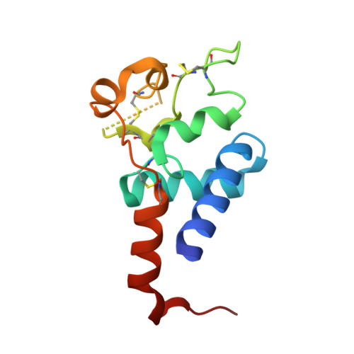 | |
UniProt & NIH Common Fund Data Resources | |||||
PHAROS: Q9H1Z9 GTEx: ENSG00000182612 | |||||
Entity Groups | |||||
| Sequence Clusters | 30% Identity50% Identity70% Identity90% Identity95% Identity100% Identity | ||||
| UniProt Group | Q9H1Z9 | ||||
Sequence AnnotationsExpand | |||||
Reference Sequence | |||||
| Length ( Å ) | Angle ( ˚ ) |
|---|---|
| a = 62.3 | α = 90 |
| b = 62.3 | β = 90 |
| c = 117.48 | γ = 90 |
| Software Name | Purpose |
|---|---|
| PHENIX | refinement |
| MxCuBE | data collection |
| XDS | data reduction |
| XSCALE | data scaling |
| PHASER | phasing |
| Coot | model building |
| Funding Organization | Location | Grant Number |
|---|---|---|
| Not funded | -- |


