9HPJ | pdb_00009hpj

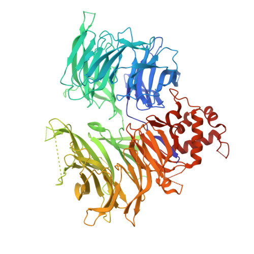
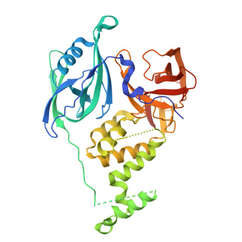
Cryo-EM structure of DDB1dB-CRBN-MRT-0031619, conformation 2


Experimental Data Snapshot

  • Method: ELECTRON MICROSCOPY
  • Resolution: 3.10 Å
  • Aggregation State: PARTICLE 
  • Reconstruction Method: SINGLE PARTICLE 

Starting Model: experimental
View more details

wwPDB Validation 3D Report Full Report

Validation slider image for 9HPJ

This is version 1.0 of the entry. See complete history. 

Literature

A degron-mimicking molecular glue drives CRBN homo-dimerization and degradation.

Langousis, G., Gainza, P., Hunkeler, M., Kapsitidou, D., Donckele, E.J., Annunziato, S., Wiedmer, L., Jones, K.F.M., DeMarco, B., Quan, C., Bunker, R.D., Lumb, K.J., Fasching, B., Castle, J.C., Townson, S.A., Bonenfant, D.

(2025) Nat Commun 16: 10157-10157

  • DOI: https://doi.org/10.1038/s41467-025-65094-3
  • Primary Citation Related Structures: 
    9HPI, 9HPJ

  • PubMed Abstract: 

    Cereblon (CRBN) is an E3 ubiquitin ligase widely harnessed for targeted protein degradation (TPD). We report the discovery of a molecular glue degrader (MGD), MRT-31619, that drives homo-dimerization of CRBN and promotes its fast, potent, and selective degradation by the ubiquitin proteasome system. Interestingly, the cryo-electron microscopy (cryo-EM) structure of the CRBN homodimer reveals a unique mechanism whereby two molecular glues assemble into a helix-like structure and drive ternary complex formation by mimicking a neosubstrate G-loop degron. This CRBN chemical knockout offers a valuable tool to elucidate the molecular mechanism of MGDs, to investigate its endogenous substrates and understand their physiological roles.


  • Organizational Affiliation: 
    • Monte Rosa Therapeutics AG, Basel, Switzerland.

Macromolecule Content 

  • Total Structure Weight: 186.95 kDa 
  • Atom Count: 12,045 
  • Modeled Residue Count: 1,499 
  • Deposited Residue Count: 1,642 
  • Unique protein chains: 2

Macromolecules

Find similar proteins by:|  3D Structure
Entity ID: 1
MoleculeChains  Sequence LengthOrganismDetailsImage
DNA damage-binding protein 1836Homo sapiensMutation(s): 0 
Gene Names: DDB1, XAP1
UniProt & NIH Common Fund Data Resources
Find proteins for Q16531 (Homo sapiens)
Explore Q16531 
Go to UniProtKB:  Q16531
PHAROS:  Q16531
GTEx:  ENSG00000167986 
Entity Groups
Sequence Clusters30% Identity50% Identity70% Identity90% Identity95% Identity100% Identity
UniProt GroupQ16531
Sequence Annotations
Expand
Reference Sequence
Find similar proteins by:|  3D Structure
Entity ID: 2
MoleculeChains  Sequence LengthOrganismDetailsImage
Protein cereblon
B, C
403Homo sapiensMutation(s): 0 
Gene Names: CRBN, AD-006
UniProt & NIH Common Fund Data Resources
Find proteins for Q96SW2 (Homo sapiens)
Explore Q96SW2 
Go to UniProtKB:  Q96SW2
PHAROS:  Q96SW2
GTEx:  ENSG00000113851 
Entity Groups
Sequence Clusters30% Identity50% Identity70% Identity90% Identity95% Identity100% Identity
UniProt GroupQ96SW2
Sequence Annotations
Expand
Reference Sequence

Experimental Data & Validation

Experimental Data

  • Method: ELECTRON MICROSCOPY
  • Resolution: 3.10 Å
  • Aggregation State: PARTICLE 
  • Reconstruction Method: SINGLE PARTICLE 
EM Software:
TaskSoftware PackageVersion
RECONSTRUCTIONRELION5.0
MODEL REFINEMENTPHENIX1.21

Structure Validation

View Full Validation Report


Entry History 

& Funding Information

Deposition Data


Funding OrganizationLocationGrant Number
Not funded--

Revision History  (Full details and data files)

  • Version 1.0: 2025-12-03
    Type: Initial release