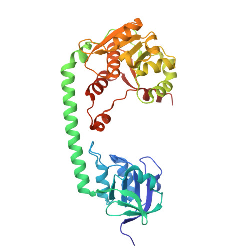
9HMZ | pdb_00009hmz

Structure of Corynebacterium glutamicum SteA, a cell division regulator


Experimental Data Snapshot

  • Method: X-RAY DIFFRACTION
  • Resolution: 2.05 Å
  • R-Value Free: 
    0.238 (Depositor), 0.222 (DCC) 
  • R-Value Work: 
    0.205 (Depositor), 0.193 (DCC) 
  • R-Value Observed: 
    0.207 (Depositor) 

Starting Model: experimental
View more details

wwPDB Validation 3D Report Full Report

Validation slider image for 9HMZ

This is version 1.1 of the entry. See complete history

Literature

Mechanistic insights into SteAB regulation of cell wall hydrolase RipA in Mycobacterium tuberculosis.

Carloni, G.Gaday, Q.Megrian, D.Petit, J.Martinez, M.Sogues, A.Ben Assaya, M.Kakonyi, M.Haouz, A.Alzari, P.M.Wehenkel, A.M.

(2026) mBio 17: e0370025-e0370025

  • DOI: https://doi.org/10.1128/mbio.03700-25
  • Primary Citation Related Structures: 
    9HLE, 9HMX, 9HMY, 9HMZ

  • PubMed Abstract: 

    D,L-endopeptidase RipA is the major peptidoglycan (PG) hydrolase required for cell separation in Mycobacterium tuberculosis ( Mtb ), as RipA defects severely hinder cell division and increase antibiotic vulnerability. Despite extensive studies, the mechanisms governing Mtb RipA regulation remain controversial and poorly understood. Here, we report an integrative structural and functional analysis of the Mtb SteAB system, a regulatory transmembrane septal complex encoded by adjacent genes Rv1697 ( steA ) and Rv1698/mctB ( steB ) that are conserved across Mycobacteriales . The separate crystal structures of the cytoplasmic core of Mt SteA and the periplasmic core of Mt SteB in complex with the RipA coiled-coil domain (RipA CC ), along with biochemical evidence that these proteins form a stable transmembrane physical complex, define the function of the SteAB complex as a regulator of RipA-mediated cell separation in Mtb , arguing against a previously proposed role of MctB (Rv1698) as a putative mycobacterial outer membrane copper transporter. Our structural findings showed that, upon Mt SteB binding, RipA CC is oriented perpendicular to the membrane, bringing its endopeptidase catalytic domain in physical contact with the PG layer, while the homodimeric structural core of SteA revealed a conserved functional pocket similar to the phosphonucleotide-binding site of thiamine pyrophosphokinase. These data, coupled with the in vivo phenotypic analysis of a steAB knockout mutant of Cglu , support a model in which the SteAB heterotetramer orchestrates the productive positioning of RipA leading to PG hydrolysis activation. These findings shed new light on the regulation of mycobacterial cell wall remodeling, with implications for understanding Mtb pathogenesis and identifying novel antimicrobial targets.IMPORTANCEPeptidoglycan (PG) is a major component of the bacterial cell wall. A flexible but strong PG mesh encloses the cell, conferring mechanical resistance and preventing cell lysis. This PG mesh is continually remodeled during the bacterial life by the coordinated action of tightly regulated PG-hydrolases and synthetases. Here, we report the structural characterization of the M. tuberculosis SteAB system, which regulates the action of the major enzyme responsible for disassembling the PG mesh to allow daughter cell separation at the end of cell division. The proposed model for the septal control of PG hydrolysis illustrates how the transmembrane SteAB complex can promote enzyme activation and provides structural information that may help target the activation mechanism for antibiotic development.


  • Organizational Affiliation
    • Institut Pasteur, Université Paris Cité, CNRS UMR 3528, Bacterial Cell Cycle Mechanisms Unit, Paris, France.

Macromolecule Content 

  • Total Structure Weight: 280.54 kDa 
  • Atom Count: 20,802 
  • Modeled Residue Count: 2,635 
  • Deposited Residue Count: 2,664 
  • Unique protein chains: 1

Macromolecules

Find similar proteins by:|  3D Structure
Entity ID: 1
MoleculeChains  Sequence LengthOrganismDetailsImage
SteA-like C-terminal domain-containing protein
A, B, C, D, E
A, B, C, D, E, F, G, H
333Corynebacterium glutamicum ATCC 13032Mutation(s): 0 
Gene Names: Cgl1415
UniProt
Find proteins for Q8NQL9 (Corynebacterium glutamicum (strain ATCC 13032 / DSM 20300 / JCM 1318 / BCRC 11384 / CCUG 27702 / LMG 3730 / NBRC 12168 / NCIMB 10025 / NRRL B-2784 / 534))
Explore Q8NQL9 
Go to UniProtKB:  Q8NQL9
Entity Groups
Sequence Clusters30% Identity50% Identity70% Identity90% Identity95% Identity100% Identity
UniProt GroupQ8NQL9
Sequence Annotations
Expand
Reference Sequence

Experimental Data & Validation

Experimental Data

  • Method: X-RAY DIFFRACTION
  • Resolution: 2.05 Å
  • R-Value Free:  0.238 (Depositor), 0.222 (DCC) 
  • R-Value Work:  0.205 (Depositor), 0.193 (DCC) 
  • R-Value Observed: 0.207 (Depositor) 
Space Group: P 21 21 21
Unit Cell:
Length ( Å )Angle ( ˚ )
a = 126.506α = 90
b = 128.72β = 90
c = 211.85γ = 90
Software Package:
Software NamePurpose
autoPROCdata processing
XDSdata reduction
STARANISOdata scaling
PHASERphasing
BUSTERrefinement
Cootmodel building

Structure Validation

View Full Validation Report


Entry History 

& Funding Information

Deposition Data


Funding OrganizationLocationGrant Number
Agence Nationale de la Recherche (ANR)FranceANR-21-CE11-0003

Revision History  (Full details and data files)

  • Version 1.0: 2025-07-30
    Type: Initial release
  • Version 1.1: 2026-09-02
    Changes: Database references