Organocatalytic Switches of DNA Glycosylase OGG1 Catalyze a Highly Efficient AP-Lyase Function.
Kehler, M., Zhou, K., Kemas, A.M., Del Prado, A., Hutchinson, E.S., Nairn, E.H., Varga, M., Plattner, Y., Zhong, Y., Purewal-Sidhu, O., Haslam, J., Wiita, E., Gildie, H., Singerova, K., Szaruga, Z., Almlof, I., Hormann, F.M., Liu, K.C., Wallner, O., Ortis, F., Homan, E.J., Gileadi, O., Rudd, S.G., Stenmark, P., de Vega, M., Helleday, T., D'Arcy-Evans, N.D., Lauschke, V.M., Michel, M.(2025) Chemistry 31: e202500382-e202500382
- PubMed: 40294343 Search on PubMed
- DOI: https://doi.org/10.1002/chem.202500382
- Primary Citation Related Structures:
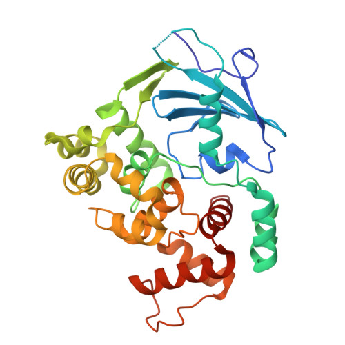
9HLF - PubMed Abstract:
8-oxoGuanine DNA glycosylase 1 (OGG1) is the first known target of organocatalytic switches (ORCAs), which rewrite the biochemical function of the enzyme through redirection of its preferred substrate from 8-oxoG to AP sites. Previously, different ORCA chemotypes were shown to enhance the operational pH window for OGG1, possibly through direct involvement in proton transfer events during DNA strand cleavage. Accordingly, compound pK a is a crucial and necessary consideration for the identification and application of future OGG1 ORCAs. Here, we identify a minimal structure of organocatalytic switches-4-anilino pyridines and 6-anilino pyrimidines-which are dimethyl-amino-pyridine (DMAP)-type Brønsted bases binding the active site of OGG1. Systematic interrogation of compound basicity through modulation of electron-withdrawing (EWG) and electron-donating (EDG) substituents reveals that a pK a less or equal to the assay pH is a viable parameter for prediction of compound activity. The lead structure (AC 50 13 nM, pK a 7.0) was then identified as a potent scaffold from a screen in a patient-derived 3D model of metabolic dysfunction-associated steatohepatitis (MASH), where it reduced hepatic fibrosis by 35%. Collectively, these findings deepen the knowledge of this novel modulator class, with important implications for future enzyme targets and probe development.
- Science for Life Laboratory, Department of Oncology-Pathology, Karolinska Institutet, Stockholm, Sweden.
Organizational Affiliation: