9HJP | pdb_00009hjp

Improved structure of mouse Gasdermin D


Experimental Data Snapshot

  • Method: X-RAY DIFFRACTION
  • Resolution: 2.64 Å
  • R-Value Free: 
    0.260 (Depositor), 0.274 (DCC) 
  • R-Value Work: 
    0.239 (Depositor), 0.237 (DCC) 
  • R-Value Observed: 
    0.240 (Depositor) 

Starting Model: experimental
View more details

wwPDB Validation 3D Report Full Report

Validation slider image for 9HJP

This is version 1.1 of the entry. See complete history

Literature

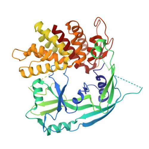
Improved structure of mouse gasdermin D: a new blueprint for structure-based drug design.

De Colibus, L.Ludzia, P.Biasutto, A.Pica, A.Hopper, J.T.S.Jazayeri, A.Durr, K.L.

(2025) Acta Crystallogr F Struct Biol Commun 81: 408-415

  • DOI: https://doi.org/10.1107/S2053230X25007149
  • Primary Citation Related Structures: 
    9HJP

  • PubMed Abstract: 

    Gasdermin D (GSDMD) is a protein that has gained significant attention in recent years due to its crucial role in inflammatory cell death, particularly pyroptosis. Pyroptosis is a highly inflammatory form of programmed cell death that is triggered by various microbial infections and sterile inflammatory stimuli. GSDMD acts as an executioner molecule in this process, leading to the release of pro-inflammatory cytokines and amplifying the immune response. Here, we present a higher resolution, significantly improved apo crystal structure of the deposited mouse structure model that will be beneficial for structure-based drug-design approaches towards this important pharmacological target.


  • Organizational Affiliation
    • OMass Therapeutics, Building 4000, Chancellor Court, John Smith Drive, Oxford Business Park, ARC, Oxford OX4 2GX, United Kingdom.

Macromolecule Content 

  • Total Structure Weight: 99.85 kDa 
  • Atom Count: 6,217 
  • Modeled Residue Count: 790 
  • Deposited Residue Count: 904 
  • Unique protein chains: 1

Macromolecules

Find similar proteins by:|  3D Structure
Entity ID: 1
MoleculeChains  Sequence LengthOrganismDetailsImage
Gasdermin-D
A, B
452Mus musculusMutation(s): 0 
Gene Names: GsdmdGsdmdc1
UniProt & NIH Common Fund Data Resources
Find proteins for Q9D8T2 (Mus musculus)
Explore Q9D8T2 
Go to UniProtKB:  Q9D8T2
IMPC:  MGI:1916396
Entity Groups
Sequence Clusters30% Identity50% Identity70% Identity90% Identity95% Identity100% Identity
UniProt GroupQ9D8T2
Sequence Annotations
Expand
Reference Sequence

Small Molecules

Ligands 3 Unique
IDChains Name / Formula / InChI Key2D Diagram3D Interactions
PE8

Query on PE8


Download:Ideal Coordinates CCD File
D [auth B]3,6,9,12,15,18,21-HEPTAOXATRICOSANE-1,23-DIOL
C16 H34 O9
GLZWNFNQMJAZGY-UHFFFAOYSA-N
CL

Query on CL


Download:Ideal Coordinates CCD File
C [auth A],
E [auth B],
G [auth B]
CHLORIDE ION
Cl
VEXZGXHMUGYJMC-UHFFFAOYSA-M
NA

Query on NA


Download:Ideal Coordinates CCD File
F [auth B],
H [auth B],
I [auth B]
SODIUM ION
Na
FKNQFGJONOIPTF-UHFFFAOYSA-N

Experimental Data & Validation

Experimental Data

  • Method: X-RAY DIFFRACTION
  • Resolution: 2.64 Å
  • R-Value Free:  0.260 (Depositor), 0.274 (DCC) 
  • R-Value Work:  0.239 (Depositor), 0.237 (DCC) 
  • R-Value Observed: 0.240 (Depositor) 
Space Group: P 1 21 1
Unit Cell:
Length ( Å )Angle ( ˚ )
a = 67.209α = 90
b = 86.934β = 95.24
c = 82.466γ = 90
Software Package:
Software NamePurpose
BUSTERrefinement
autoPROCdata reduction
autoPROCdata scaling
MOLREPphasing

Structure Validation

View Full Validation Report


Entry History 

& Funding Information

Deposition Data


Funding OrganizationLocationGrant Number
Other privateUnited KingdomNA

Revision History  (Full details and data files)

  • Version 1.0: 2025-10-01
    Type: Initial release
  • Version 1.1: 2026-04-15
    Changes: Database references