9HD4 | pdb_00009hd4

LecB in complex with photoswitchable compound GTB


Experimental Data Snapshot

  • Method: X-RAY DIFFRACTION
  • Resolution: 1.49 Å
  • R-Value Free: 
    0.215 (Depositor), 0.216 (DCC) 
  • R-Value Work: 
    0.148 (Depositor), 0.158 (DCC) 

Starting Model: experimental
View more details

wwPDB Validation   3D Report Full Report


Ligand Structure Quality Assessment 


This is version 1.1 of the entry. See complete history


Literature

Synthesis, photochemical and biological evaluation of novel photoswitchable glycomimetic ligands of Pseudomonas aeruginosa LecB.

Bhattacharya, S.Tempra, G.Colleoni, A.Matera, C.Castagna, R.Parisini, E.

(2025) RSC Adv 15: 49796-49808

  • DOI: https://doi.org/10.1039/d5ra06897e
  • Primary Citation of Related Structures:  
    9HD4

  • PubMed Abstract: 

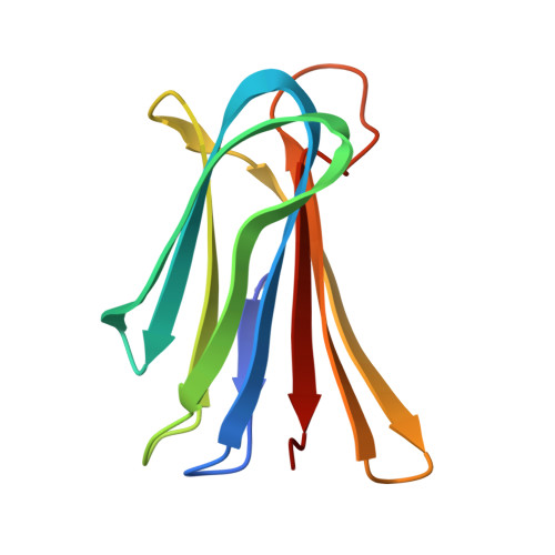
    Bacterial multidrug resistance (MDR) poses a major threat to global health. The continued use of antibiotics, combined with genetic variations and exposure to nosocomial infections, has led to the selection and spread of multidrug-resistant bacteria. In recent years, photopharmacology has emerged as a strategy to combat MDR by enabling precise, light-controlled spatiotemporal modulation of the biological activity of photo-switchable compounds. Among different microbial species, Pseudomonas aeruginosa is a prominent bacterium involved in acute and chronic lung infections, posing a significant health concern, particularly among hospitalized and immunocompromised patients. The bacterium's capacity to form biofilms, a key factor in the development of MDR, is closely linked to the activity of the virulence factor LecB, a carbohydrate-binding protein with a well-documented role in biofilm formation. In this study, we report the design, synthesis and biological evaluation of two novel photoswitchable LecB modulators, photofucose-1 and photofucose-2. Isothermal Titration Calorimetry (ITC) analysis revealed that photofucose-2 binds LecB with high affinity, exhibiting a distinct difference in dissociation constants ( K d ) between its cis and trans isomers. Moreover, we determined the X-ray crystal structure of the LecB-photofucose-2 complex, offering insights into its binding mechanism. These findings lay the groundwork for the rational, structure-based design of novel light-responsive compounds targeting LecB and represent a potential new avenue in the development of innovative strategies to combat bacterial resistance.


  • Organizational Affiliation
    • Department of Biotechnology, Latvian Institute of Organic Synthesis Aizkraukles 21 LV-1006 Riga Latvia emilio.parisini@osi.lv.

Macromolecules
Find similar proteins by:  (by identity cutoff)  |  3D Structure
Entity ID: 1
MoleculeChains Sequence LengthOrganismDetailsImage
Fucose-binding lectin PA-IIL
A, B, C, D
114Pseudomonas aeruginosaMutation(s): 0 
Gene Names: lecBPA14_20610
UniProt
Find proteins for Q9HYN5 (Pseudomonas aeruginosa (strain ATCC 15692 / DSM 22644 / CIP 104116 / JCM 14847 / LMG 12228 / 1C / PRS 101 / PAO1))
Explore Q9HYN5 
Go to UniProtKB:  Q9HYN5
Entity Groups  
Sequence Clusters30% Identity50% Identity70% Identity90% Identity95% Identity100% Identity
UniProt GroupQ9HYN5
Sequence Annotations
Expand
  • Reference Sequence
Small Molecules
Ligands 2 Unique
IDChains Name / Formula / InChI Key2D Diagram3D Interactions
A1IT5 (Subject of Investigation/LOI)
Query on A1IT5

Download Ideal Coordinates CCD File 
E [auth A],
J [auth B],
M [auth C],
P [auth D]
~{N}-[[(2~{R},3~{S},4~{R},5~{S},6~{S})-6-methyl-3,4,5-tris(oxidanyl)oxan-2-yl]methyl]-4-[(~{Z})-phenyldiazenyl]benzenesulfonamide
C19 H23 N3 O6 S
FZXKDZRYIBYABH-GHJLDVCKSA-N
CA
Query on CA

Download Ideal Coordinates CCD File 
F [auth A]
G [auth A]
H [auth A]
I [auth A]
K [auth B]
F [auth A],
G [auth A],
H [auth A],
I [auth A],
K [auth B],
L [auth B],
N [auth C],
O [auth C],
Q [auth D],
R [auth D]
CALCIUM ION
Ca
BHPQYMZQTOCNFJ-UHFFFAOYSA-N
Experimental Data & Validation

Experimental Data

  • Method: X-RAY DIFFRACTION
  • Resolution: 1.49 Å
  • R-Value Free:  0.215 (Depositor), 0.216 (DCC) 
  • R-Value Work:  0.148 (Depositor), 0.158 (DCC) 
Space Group: C 2 2 21
Unit Cell:
Length ( Å )Angle ( ˚ )
a = 64.21α = 90
b = 83.72β = 90
c = 158.88γ = 90
Software Package:
Software NamePurpose
REFMACrefinement
autoPROCdata reduction
autoPROCdata scaling
REFMACphasing

Structure Validation

View Full Validation Report


Ligand Structure Quality Assessment 


Entry History & Funding Information

Deposition Data


Funding OrganizationLocationGrant Number
European Union (EU)European Union--

Revision History  (Full details and data files)

  • Version 1.0: 2025-11-19
    Type: Initial release
  • Version 1.1: 2026-01-28
    Changes: Database references